

Platelet-rich plasmaPlatelet-rich plasma therapy in ophthalmology
What is PRP-therapy?
PRP PlateletRich Plasma) It is an autologous serum, obtained from the patient's own blood, enriched with an increased concentration of platelets. It is composed of two main components:
- Plasma is the liquid part of blood
- Platelets are blood cells that participate not only in blood clotting but also in the processes of repair and regeneration, thanks to their rich content of growth factors.
Platelets release biologically active substances such as PDGF, TGF-β, VEGF, and others, which aid in cellular repair and stimulate tissue regeneration.
Applications of PRP in ophthalmology
PRP therapy is used in the treatment of a wide range of eye diseases and conditions, including:
- Dry macular degeneration
- Glaucoma
- Dry eye syndrome
- Amblyopia (lazy eye)
- Optic nerve atrophy
- Genetic retinal diseases (e.g., Stargardt disease, retinitis pigmentosa)
- Traumatic eye injuries
- Corneal injuries and erosions
How is enhanced plasma obtained?
The procedure includes the following steps:
- Drawing venous blood from the patient.
- Centrifugation – Blood is placed in a special centrifuge, which separates the platelet-rich plasma from the other blood components.
- Plasma isolation – the obtained fraction is used for injection into the specific treatment area.
What is a PRP injection?
The PRP solution is injected directly into the affected tissue. In ophthalmology, this is most commonly performed in the periocular area. Although the exact mechanism of action is still being investigated, clinical observations and studies indicate that the concentrated growth factors in PRP accelerate restorative processes in the eye.
Platelet-rich plasma (PRP) contains not only platelets but also white blood cells, stem cells, bioactive proteins, cytokines, and other regenerative factors. This makes it extremely effective in treating eye tissues, as it helps create a biological „scaffold“ for repair and healing.
Since PRP is derived from the patient's own blood, the risk of immunological reactions is eliminated, ensuring a completely biocompatible form of treatment.
Procedure for administering a PRP injection
- Primary eye examination – includes visual field, OCT, and other necessary tests.
- Venous blood collection and centrifugation.
- Preparation of the injection solution.
- Pain relief of the eye area with drops.
- Injecting PRP into the lower outer part of the eye.
- Applying a cold compress after the procedure.
The procedure is painless and generally takes about 90–120 minutes. After injection, the patient may feel mild swelling or a foreign body sensation in the eye, which subsides within a few hours to the next day.
Treatment and follow-up plan
- A follow-up check is performed two weeks after the first injection.
- If there is a positive response, two more injections are given at two-week intervals, i.e., a total of three injections for the initial course and to achieve optimal results.
- The patient is then monitored individually – in case of a new decrease in vision, a maintenance injection may be administered after 3, 6, 12 months or more, depending on their condition.
- If there is no result after the first injection, the treatment is discontinued.
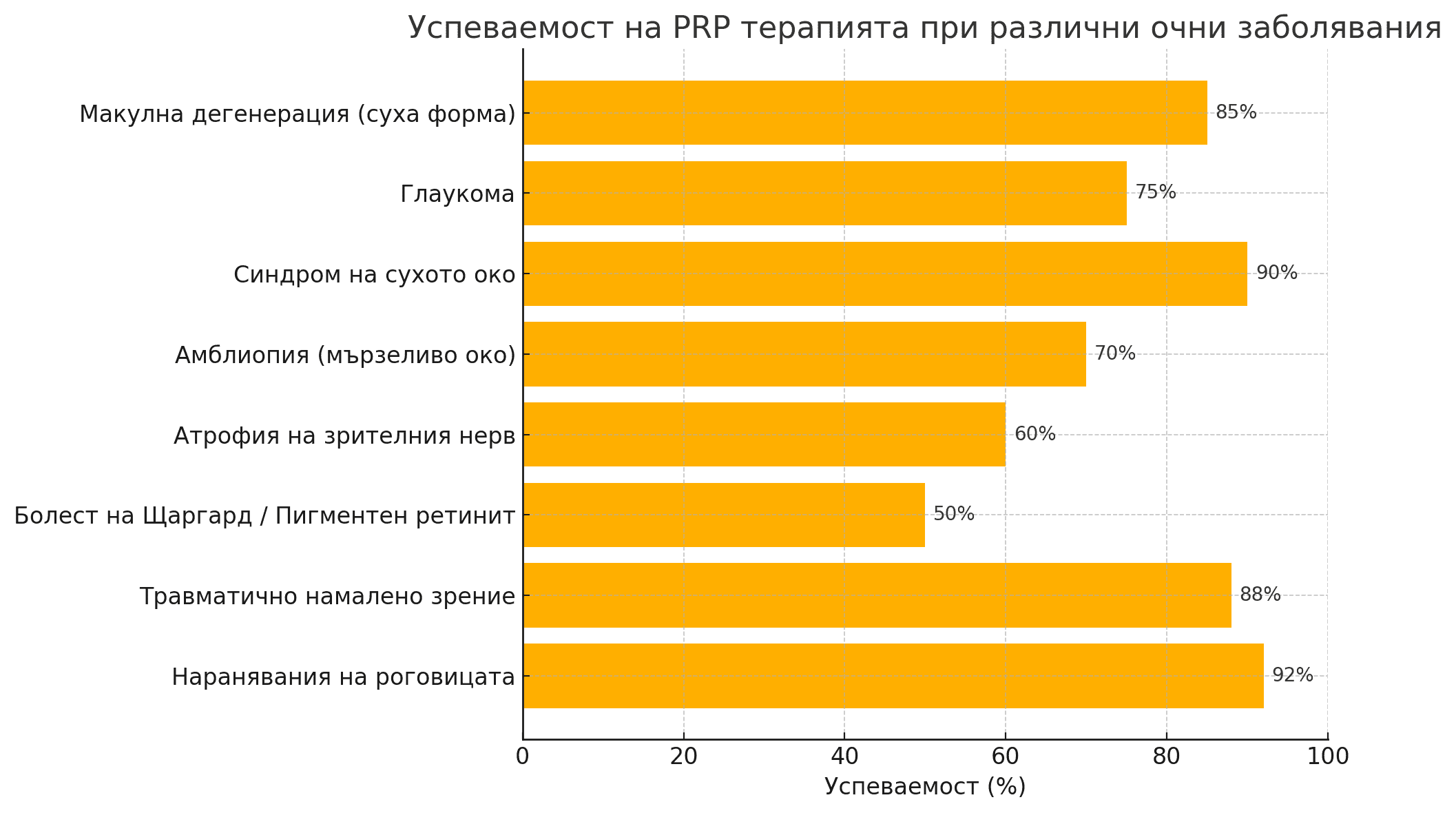
About 80–90% of the patients show a positive response to therapy.
PRP therapy - risks and side effects
PRP therapy is considered low-risk because it uses the patient's own biological materials. Potential, though rare, side effects include:
- Light bleeding
- Infection
- Tissue damage
- Nerve structure damage
PRP therapy is an innovative, biological, and safe method for treating various ophthalmological diseases. Thanks to its regenerative potential, it offers new possibilities for conditions where conventional medicine has limited effectiveness. An individualized approach to each patient is key to achieving optimal results.