[everest_counter id='11512′]
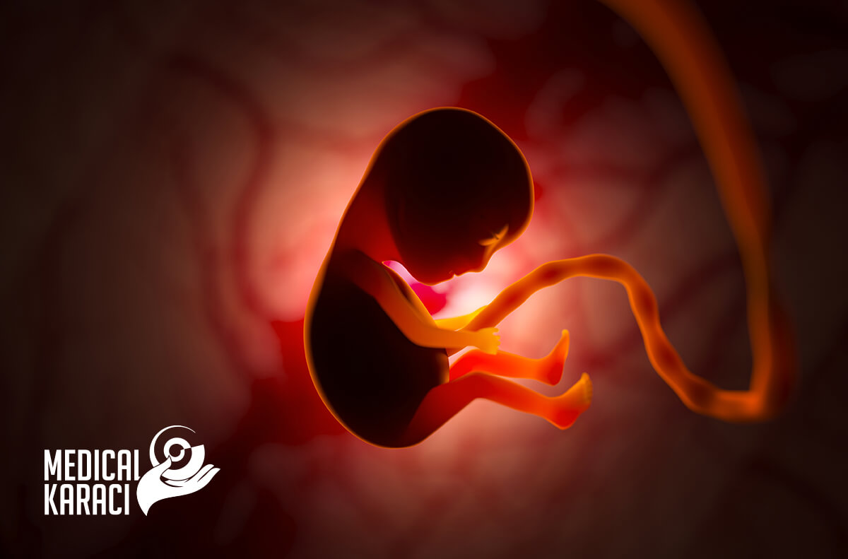
Prenatal surgery, also known as fetal surgery or antenatal surgery, is a relatively young field in medicine. Fetal surgery encompasses a wide range of surgical techniques that are performed on the unborn baby (fetus) in the uterus (in utero) to aid in proper fetal development. Early intervention before the baby is born can treat life-threatening birth defects and improve intrauterine conditions. Advances in technology allow earlier and more accurate diagnosis of diseases and birth problems in the fetus. In the last 20 years, the fetus has come to be seen as a patient, giving rise to a range of screening tests to assess intrauterine development and enabling the application of intrauterine fetal surgery to a range of fetal structural defects
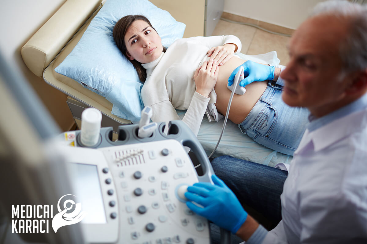
Pregnancy tracking requires three basic examinations by a fetal medicine specialist:
- First trimester: 11 - 13th gestational week
Testing in the first trimester of pregnancy includes fetal ultrasound and blood tests. The risk of aneuploidy is assessed. Examination in this period aims to evaluate the nuchal translucency, nasal bone, tricuspid valve and ductus venosus of the fetus. The combination of the examinations and the mother's age are entered into a special computer program that calculates the individual risk for Down syndrome, Edwards syndrome, Patau syndrome, and monosomy X. The sensitivity of the test is about 95%
- Second trimester: 19th - 23rd gestational week
In the second trimester of pregnancy, ultrasound examination and biochemical blood tests are performed. It is done to assess the risk of Down syndrome, other chromosomal abnormalities such as Trisomy 18 and neural tube defects- spina bifida and certain brain abnormalities. The presence of structural malformations is subject to consultation with a specialist in fetal surgery and fetal medicine.
- Third trimester: 32 - 34th gestational week
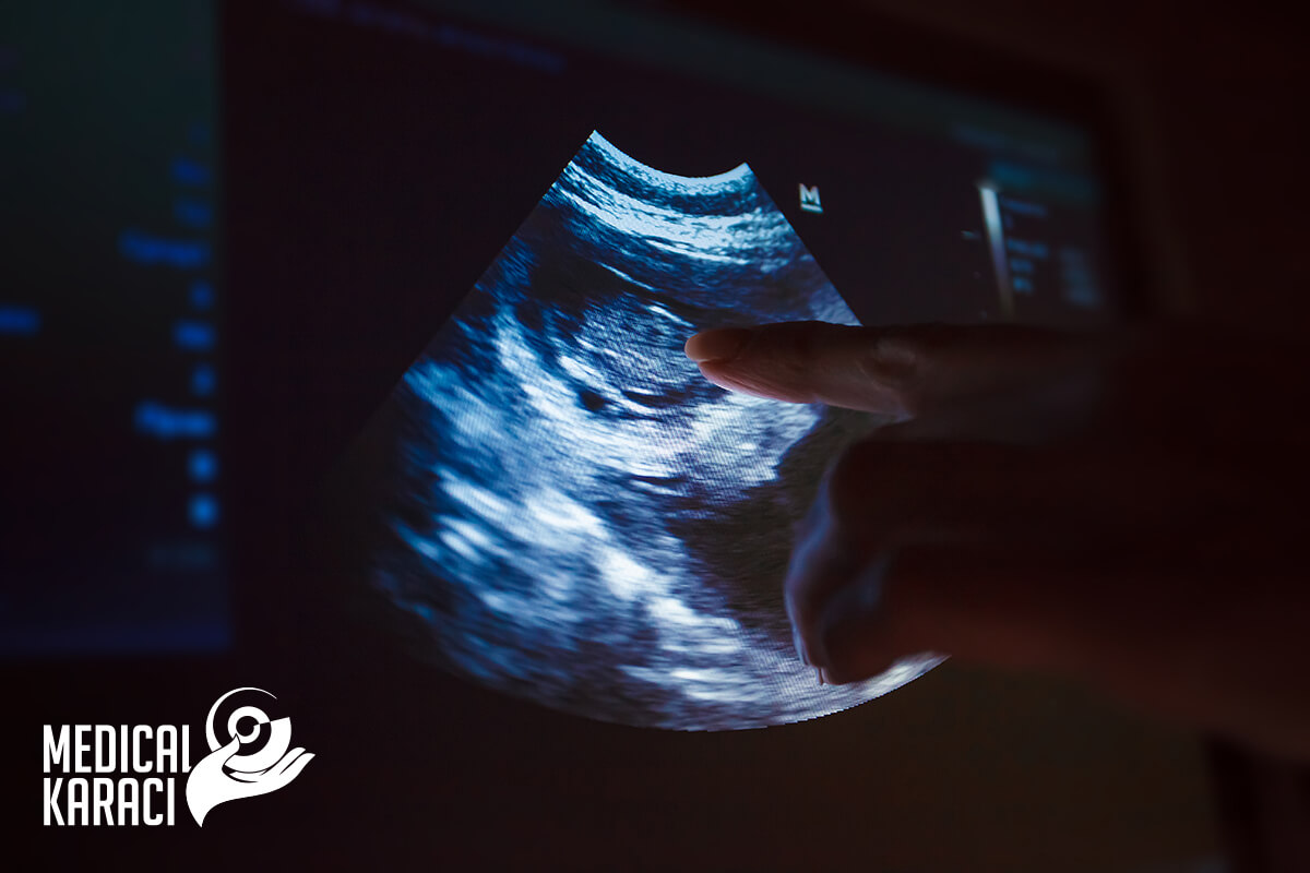
Ultrasound examination in the third trimester gives an estimate of the condition of the fetus. Doppler ultrasound provides information about blood flow in the umbilical cord vessels, middle cerebral artery, and ductus venosus. The examination also verifies the fetal anatomy. The various organs and systems of the fetus are evaluated.

When is fetal surgery applied?
With the help of fetal surgery, many birth defects in the womb can be treated, including:
- Amniotic band syndrome
- Bronchopulmonary sequestration of the lung
- Congenital cystic adenomatoid malformation (CCAM) of the lung
- Congenital diaphragmatic hernia (CDH)
- Congenital high airway obstruction syndrome (CHAOS)
- Fetal anemia
- Lower urinary tract obstruction (LUTO)
- Mediastinal teratoma
- Sacrococcygeal teratoma (SCT)
- Spina bifida (myelomeningocele)
- Double anemia-polycythemia (TAPS)
- Double reverse arterial perfusion sequence (TRAP)
- Transfusion syndrome (TTTS)
Techniques of fetal surgery
Fetal surgery uses a multidisciplinary approach to diagnose and treat a range of fetal health conditions or complications that develop during pregnancy. This type of surgery requires experience and state-of-the-art technology to provide the best possible outcomes for both the mother and her unborn baby.
The main method of prenatal surgery is open fetal surgery.

What is open fetal surgery?
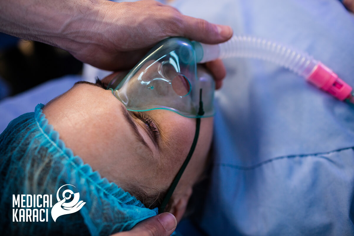
In open fetal surgery, the mother is anesthetized. Anaesthesia relaxes the uterus and allows the placenta to continue to supply oxygen to the baby. Anesthesia also passes through the placenta to the unborn child, allowing fetal surgeons to perform necessary procedures without causing pain or discomfort to the baby. An incision is made in the lower abdomen to expose the uterus. The uterus is opened using a special device to prevent bleeding. Warmed fluids are continuously infused into the uterus to keep the amniotic fluid level safe for the mother and baby. Fetal surgery is performed inside the womb and then the uterus is closed.
Open fetal surgery is a major abdominal operation like a caesarean section, except that at the end of the operation the mother is still pregnant. The intervention requires hospitalization for 3 to 7 days, requires cesarean delivery of this and future pregnancies, and often causes preterm delivery. Pregnancy should be carefully monitored by a specialist. The intake of medication to control preterm labour will also be necessary.
Fetal surgery would not be possible without advances in prenatal imaging, improved anesthesia techniques, improved surgical techniques, and physicians' knowledge and experience of prenatal complications and fetal diseases.
When is open fetal surgery recommended?
- Operation on spina bifida
Open fetal surgery for overt spinal defects (myelomeningocele and myeloschisis) requires closure of the fetal skin and tissue over the developing spinal cord early in pregnancy. Research has shown that intrauterine fetal repair reduces the need for shunt placement after birth and improves movement by preventing further neuronal damage and cerebrospinal fluid leakage.
- Removal of large congenital airway lesions
Congenital pulmonary airway malformation is a benign (non-cancerous) congenital cystic lung lesion or mass that forms in the lower airways of an unborn baby during development. It appears during the early stages of lung fetal development, but grows rapidly during the second trimester (around 20-28 gestational weeks).
Before making the choice of open fetal surgery to treat these lung lesions, two courses of steroid medication may be administered to determine if this can stop the growth. If the mass continues to grow despite steroids, open fetal surgery may be considered if there is a risk of fetal hydrops and the pregnancy is before 32 weeks gestational age.
- Removal of large sacrococcygeal teratomas
Sacrococcygeal teratoma (SCT) is one of the most common congenital germ cell tumors. It is a mass located at the base of the tailbone and occurs in approximately one in every 35,000 births.
In these cases, open fetal surgery is suggested to allow controlled removal of the tumor before birth. The operation is intended to avoid rupture of the teratoma capsule, which results in significant bleeding and can put both mother and baby at risk.
Childbirth after open fetal surgery
Since the surgical incision of the uterus in open fetal surgery does not heal as well as in caesarean section normal delivery should be avoided in the present and future pregnancies. All future births should be by planned caesarean section.
Other techniques of prenatal surgery:
Fetoscopy
Fetoscopy is an endoscopic procedure that allows surgical access to the fetus, amniotic cavity, umbilical cord, and fetal side of the placenta during pregnancy. A small (3-4 mm) incision is made on the abdomen and an endoscope is inserted through the abdominal wall and uterus into the amniotic cavity. Ultrasound technology helps guide the fetoscope into the uterus. Fetoscopy allows medical interventions such as biopsy (tissue sample), laser occlusion of abnormal blood vessels (such as chorioangioma) or treatment of spina bifida.
Fetoscopy is usually performed in the second or third trimester of pregnancy. Fetoscopic surgery is much less invasive than open fetal surgery, thus reducing the risk of preterm birth. During the procedure, mothers are given anesthesia to help control pain and anxiety. The fetus is also given medication to reduce movement and prevent pain.
Because the uterine incision for fetoscopic surgery is very small, it often heals well. Mothers can give birth naturally in current and future pregnancies.
Fetal laser surgery
Fetal laser surgeries are used to treat and correct life-threatening congenital conditions that affect monochorionic twins - when identical twins (or multiple) share a placenta - as in transfusion syndrome.
Laser surgery for twin transfusion syndrome-fetoscopic laser ablation
What is Selective Fetoscopic Laser Photocoagulation (SFLP)?
Selective fetoscopic laser photocoagulation (SFLP), also called fetoscopic selective laser ablation, is a minimally invasive procedure performed on the placenta of infants with transfusion syndrome. Using a fetoscope, the pathological connections of the placental blood vessels between the babies are detected and then sealed using laser energy. Laser ablation stops the abnormal fluid exchange between the twins.
Which conditions are treated with SFLP?
In severe cases of transfusion syndrome in twins, laser surgery is usually the best treatment option.
Why is surgery necessary for transfusion syndrome?
A fetus diagnosed with TTTS must undergo SFLP in order for the disease and its complications to be stopped or reversed. This disease occurs because the twins share a placenta (monochorionic) and there are connections between the blood vessels of the placenta, allowing the twins to share fluids with each other. Thus, one baby develops as a "donor" and the other is a "recipient" of placental blood. If this abnormal sharing is significant, the twins can become ill and face severe complications including: heart failure, brain damage and death. Transfusion syndrome occurs in one in five pregnancies in identical twins. The condition threatens the lives of both babies.
Laser surgery is performed to seal abnormal blood vessel connections, stop harmful fluid sharing, and correct or prevent complications. In the most severe cases of double transfusion, survival rates can be as low as 10 to 15% if left untreated.
What happens during selective femtoscopic laser photocoagulation?
Before laser surgery for transfusion syndrome, an anesthesiologist places an epidural to make you comfortable during the procedure. Once you are comfortable and pain-free, the procedure begins. The fetoscope is inserted through a small incision in the amniotic cavity. The scope allows the abnormal fetal connections between blood vessels to be clearly seen and identified and closed. A laser is then used to seal these connections.
Once the laser procedure is complete, amnioreduction is usually performed to remove excess amniotic fluid. This is done through the same fetoscope.
After the procedure, a day of hospitalization is required to keep you under observation for signs of contractions or complications.
What is the recovery after laser ablation?
SFLP is usually not associated with significant recovery time and there is only minimal pain. Reduced activity is recommended throughout the remainder of pregnancy given the increased risk of preterm delivery.
Amniocentesis
Amniocentesis is a test that may be offered to you during pregnancy to check whether your baby has a genetic or chromosomal mutation, such as Down syndrome, Edwards syndrome or Patau syndrome. It involves taking and testing a small sample of cells from the amniotic fluid, or the fluid that surrounds the unborn baby in the womb (uterus).
When is amniocentesis offered?
Amniocentesis is not offered to all pregnant women. It is only offered if there is a greater risk that your baby will have a genetic condition.
This can happen if:
- An antenatal screening test suggests that your baby may be born with a mutation, such as Down syndrome, Edwards syndrome or Patau syndrome
- you have had a previous pregnancy that was affected by a genetic disorder
- you have a family history of a genetic disease, such as sickle cell disease, thalassaemia, cystic fibrosis or muscular dystrophy.
It is important to remember that it is not mandatory to do amniocentesis. It is up to you to make the decision after being informed in detail about the benefits and risks of the procedure.
How is amniocentesis performed?
Amniocentesis is usually performed between the 15th and 20th gestational week of pregnancy, but you can have it later if necessary. It can also be done earlier, but this may increase the risk of complications.
During the test, a long thin needle guided by an ultrasound image is inserted through the abdominal wall. The needle is passed into the amniotic sac, which surrounds the fetus, and a small sample of amniotic fluid is taken for analysis. The test itself usually takes about 10 minutes.
Amniocentesis is usually described as uncomfortable rather than painful. Some women describe experiencing pain similar to the pain during a period or feeling pressure.
The first test results should be available within 3 days. You will be notified if Down syndrome, Edwards syndrome or Patau syndrome has been detected. If rarer conditions are being tested, it may take 3 weeks or longer for the results to come out.
If your test shows that your baby has a genetic or chromosomal condition, the implications will be discussed in detail with you.
There is no cure for most conditions identified by amniocentesis, so you will need to consider your options carefully.
You can choose to continue with your pregnancy while gathering information about the condition so that you are fully prepared.
Or you might consider terminating the pregnancy.
What are the risks of amniocentesis?
Before you decide to have an amniocentesis, the risks and possible complications will be discussed with you.
One of the main risks associated with amniocentesis is miscarriage. It is estimated that this occurs in up to 1 in every 100 women who choose to have an amniocentesis.
There are also some other risks, such as infection or the need for a repeat procedure if the results are unclear.
What are the alternatives?
An alternative to amniocentesis is a test called chorionic villus sampling (CVS). This is where a small sample of cells from the placenta is removed for testing. It is usually carried out between the 11th and 14th week of pregnancy, although it can be carried out later than this if necessary. With CVS, the risk of miscarriage is similar to the risk of miscarriage with amniocentesis (up to 1 in 100). Because the test can be done earlier, you will have more time to consider the results.
Bipolar umbilical coagulation
Umbilical cord coagulation is a procedure used in twin pregnancies with a common placenta (monochorionic) to selectively terminate the life of one twin. It is a minimally invasive procedure in which a needle is guided via ultrasound into the umbilical cord and blood flow is coagulated either by radiofrequency ablation or bipolar cauterization. This interrupts blood communication between the fetuses and allows the remaining twin to progress as a single without complications. The lost baby remains in the amniotic cavity and stops developing.
Although this is a difficult decision for the family, it may be the right decision as it increases the chances for the surviving twin by eliminating the risk of death or brain damage.
How is it done?
Coagulation of the umbilical cord can be accomplished by either forceps or radiofrequency ablation. The technique depends on the specific case.
Bipolar coagulation is performed in the operating room under spinal anesthesia. A small incision is made in the skin so that a very small camera (a phetoscope) can be inserted into the uterus. Through this chamber, a bipolar forceps is directed to the umbilical cord of the affected twin. The umbilical cord is tightened to stop blood flowing to this twin. This operation takes approximately 30 to 60 minutes.
Radiofrequency ablation is performed under local anesthesia (lidocaine) in the fetal medicine office. A small needle is guided to the umbilical cord of the affected twin via ultrasound. The umbilical cord is heated so that the blood vessels in the cord are sealed. This procedure takes approximately 15 minutes.
When are these procedures applied?
The procedures are applied in monochorionic double pregnancies with one or more of the following situations:
- Severe transfusion syndrome
- Double reverse arterial perfusion
- Severe non-conforming abnormalities
- Severe selective intrauterine growth restriction
Cordocentesis

Cordocentesis - also known as cord blood sampling - is a diagnostic prenatal test in which a sample of the baby's blood is removed from the umbilical cord for testing.
Cordocentesis, which is usually done after 18 weeks of pregnancy, can be used to detect certain genetic disorders, blood conditions and infections. Cordocentesis can also be used to deliver blood and medications to the baby through the umbilical cord.
Why is it done?
Cordocentesis is primarily used to detect and treat blood conditions such as fetal anemia - a small amount of healthy red blood cells in a developing baby. Cordocentesis is usually done when the diagnosis cannot be made by amniocentesis, chorionic villus sampling, ultrasound or other methods. Cordocentesis carries a higher risk of complications for the baby, including death, than other procedures. Your doctor will only suggest the procedure if other options are not available or they will not produce results quickly enough.
Rarely, cordocentesis can be used to check fetal chromosomes by chromosome microarray or karyotype analysis. Blood obtained by cordocentesis can potentially be used for other types of genetic studies.
Risks:
Cordocentesis poses potentially serious risks, including:
- Bleeding from the fetus. Bleeding from the area where the needle was inserted is the most common complication.
- Hematoma of the umbilical cord. During or after cordocentesis, collection of fetal blood in the umbilical cord may occur. Most babies have no signs or symptoms when this happens. However, some may develop a low heart rate for a short period. If the hematoma is stable, your doctor will monitor the baby. If the hematoma is not stable or if your baby's heart rate does not recover, your doctor will recommend an emergency C-section.
- Slowing of the baby's pulse. The baby's heart rate may temporarily slow after cordocentesis.
- Infection. Rarely, cordocentesis can lead to infection of the uterus or fetus.
- Fetal-uterine bleeding. Fetal blood can enter the maternal circulation at about 40% from procedures. The amount of blood is usually small. This problem is more common when the placenta lies in the anterior uterus.
- Passage of maternal infection into the fetus. If the mother has certain infections, such as hepatitis B, hepatitis C or HIV, these can be passed on to the baby.
- Pregnancy loss. Cordocentesis carries a higher risk of fetal death than other prenatal diagnostic tests, such as chorionic villus sampling and amniocentesis. The risk is about 1 to 2% for a fetus that appears normal and is tested for genetic disorders.
Ultimately, the decision to cordocentesis is up to you. Your doctor along with a genetic specialist can help you weigh the risks and benefits of the procedure.
What can you expect?
Before the 23rd week of pregnancy, cordocentesis is usually done in an outpatient facility or doctor's office. After the 23rd week of pregnancy, cordocentesis is usually done in a hospital in case the baby develops complications that may require emergency delivery.
Before the procedure, a sample of your blood will be taken for comparison with the fetal blood samples.
During the procedure:
About 30 to 60 minutes before the procedure, you may be given antibiotics to reduce the risk of uterine infection.
Your doctor will use ultrasound to determine the location of the umbilical cord in the uterus. You will lie on your back on the table and the doctor will apply a special gel to your abdomen. The doctor will then use a small device known as an ultrasound transducer to view your baby's position on a monitor. He will then clean your tummy. Sometimes medicine is given to prevent discomfort during the procedure, but often this is not necessary.
Guided by the ultrasound, your doctor will insert a thin, hollow needle through the abdominal wall, into the uterus. A small amount of blood from the vein in the umbilical cord will be drawn into a syringe and the needle removed. You will need to lie still until the needle is inserted and the blood drawn. You may notice a burning sensation when the needle enters your skin, and feel cramping when the needle enters the uterus.
After the procedure:
You may have cramps or mild discomfort after the blood sample is taken.
Your doctor will use ultrasound to monitor your baby's heart rate after the procedure. He may suggest resting for the rest of the day. You will probably be able to resume normal activities the next day. Contact the doctor if you have vaginal bleeding or fluid leakage.
The blood sample will be analyzed in a laboratory. Test results are usually available within a few days.
Results:
Your doctor or genetic specialist will help you understand the results of your cordocentesis.
If your baby has an infection, your doctor will explain the treatment options. If your baby has severe anaemia, he may need a blood transfusion through the umbilical cord.
If the test results show that your baby has a condition that cannot be treated, you may face important decisions - for example, whether to continue or terminate the pregnancy.
Vesicoamniotic and thoracoamniotic shunting
What is vesicoamniotic and thoracoamniotic shunting?
Some fetal abnormalities can cause fluid accumulation that can be fatal if left untreated. Inserting a small tube called a shunt while the baby is still in the womb can drain the fluid and reduce the strain on the baby's body.
Vesicoamniotic shunting is used to treat urinary tract obstructions. It allows the baby's urine to drain directly into the amniotic space. Thoracoamniotic shunting is used to treat pleural effusions, congenital malformations of the pulmonary airway, and other conditions that can squeeze the baby's heart, lungs, or kidneys.
For this procedure, the mother receives either local or local anesthesia. Under ultrasound guidance, the fetal medicine specialist inserts a needle through the mother's abdomen and into the part of the baby's body filled with excess fluid. The shunt is passed through the needle and placed in position, with the end protruding from the baby's body to allow the fluid to leak into the amniotic spaces. If necessary, the surgeon can add fluids to the amniotic sac at the same time. The mother and baby receive antibiotics to prevent infection. The procedure may need to be repeated if the shunt is displaced because of the baby's movement or other reasons.
Candidates for vesicoamniotic and thoracoamniotic shunting?
If your baby's ultrasound shows a fetal abnormality that results in life-threatening fluid accumulation, your doctor may recommend a vesicoamniotic or thoracoamniotic shunt.
For safety reasons, patients must meet the following criteria:
- The baby's condition must be severe enough to warrant fetal intervention.
- The baby must not have a genetic problem or any other serious birth defect.
- The mother must be pregnant with only one baby.
While vesicoamniotic and thoracoamniotic shunting are generally considered safe and beneficial, in rare cases these procedures can lead to serious complications. It is also possible for the shunt to be displaced as the baby grows.
How does vesicoamniotic and thoracoamniotic shunting affect the baby?
Draining excess fluid can reduce the amount of accumulated abnormal mass, reverse hydrops, and greatly improve the baby's chance of survival.
How does vesicoamniotic and thoracoamniotic shunting affect pregnancy?
Your pregnancy will be closely monitored to ensure that the shunt remains in place and no further complications develop. There is a risk of premature birth. You will be able to give birth normally unless there is a medical reason for a caesarean section. You may need to be monitored every 1-2 weeks until delivery.
Intrauterine haemotransfusion
Fetal anemia is a serious complication of pregnancy and is associated with perinatal mortality and morbidity. Intrauterine transfusion provides blood to an Rh-positive fetus when the fetal red blood cells are destroyed by Rh antibodies. A characteristic of this threatening condition is that the mother's created antibodies can attack the baby's red blood cells and destroy them. This leads to fetal anemia. Blood transfusions are performed to replace the fetal red blood cells, which are destroyed by the mother's Rh-sensitive immune system. This treatment aims to keep the fetus healthy until it is mature enough to be born.
Fetal anemia can also occur as a result of a number of viruses or in the presence of multiple pregnancy. Thanks to the possibility of intrauterine blood transfusion, such cases are treatable. Transfusions can be carried out through the fetal abdomen or, more commonly, by delivering the blood into the umbilical vein or artery. Umbilical cord transfusion is the preferred method as it allows better absorption of blood and has a higher survival rate than transfusion through the abdomen.
The mother is sedated and an ultrasound image is obtained to determine the position of the fetus and placenta. After the mother's abdomen is cleaned with an antiseptic solution, she is injected with a local anaesthetic to desensitise the abdominal area where the transfusion needle will be inserted. The fetus may be given medication to temporarily stop movement.
Ultrasound is used to guide the needle through the mother's abdomen into the fetal abdomen or umbilical cord vein.
Compatible blood type (usually type O, Rh-negative) is delivered into the umbilical cord blood vessel of the fetus.
The mother is usually given antibiotics to prevent infection. She may also be given a tocolytic drug to prevent labour, although this is unusual.
What to expect after treatment?
A short recovery period is required (approximately 1 to 3 hours). If the fetus has been given medicine to prevent movement, it may be several hours until the mother feels the fetus moving again.
Why is it done?
The sensitized maternal immune system can destroy a large amount of fetal red blood cells, causing severe fetal anemia. Intrauterine blood transfusions are performed when:
- Doppler ultrasound of the middle cerebral artery suggests anemia.
- The bilirubin result from the amniocentesis test indicated that the fetus was moderately to severely affected by Rh sensitization.
- Ultrasound shows evidence of fetal hydrops, such as swollen tissues and organs.
- Taking a blood sample from the fetus indicates that the fetus has severe anemia.
The transfusion can be done immediately.
In a severely affected fetus, transfusion is done every 1 to 4 weeks until the fetus is mature enough to be born safely.
Fetal survival after transfusion depends on the severity of the disease, the method of transfusion, and the skill of the doctor doing the procedure. In general, after intrauterine transfusion through the umbilical cord:
More than 90% of the fruits that do not have hydrops survive.
About 75% of the fetuses that have hydrops survive.
Risks:
Intrauterine transfusion can cause:
- Infection of the uterus.
- Fetal infection.
- Premature birth.
- Excessive bleeding and mixing of fetal and maternal blood.
- Leakage of amniotic fluid from the uterus.
- Death of the fetus.
For more information, you can call +359895770869.