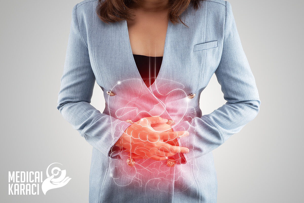
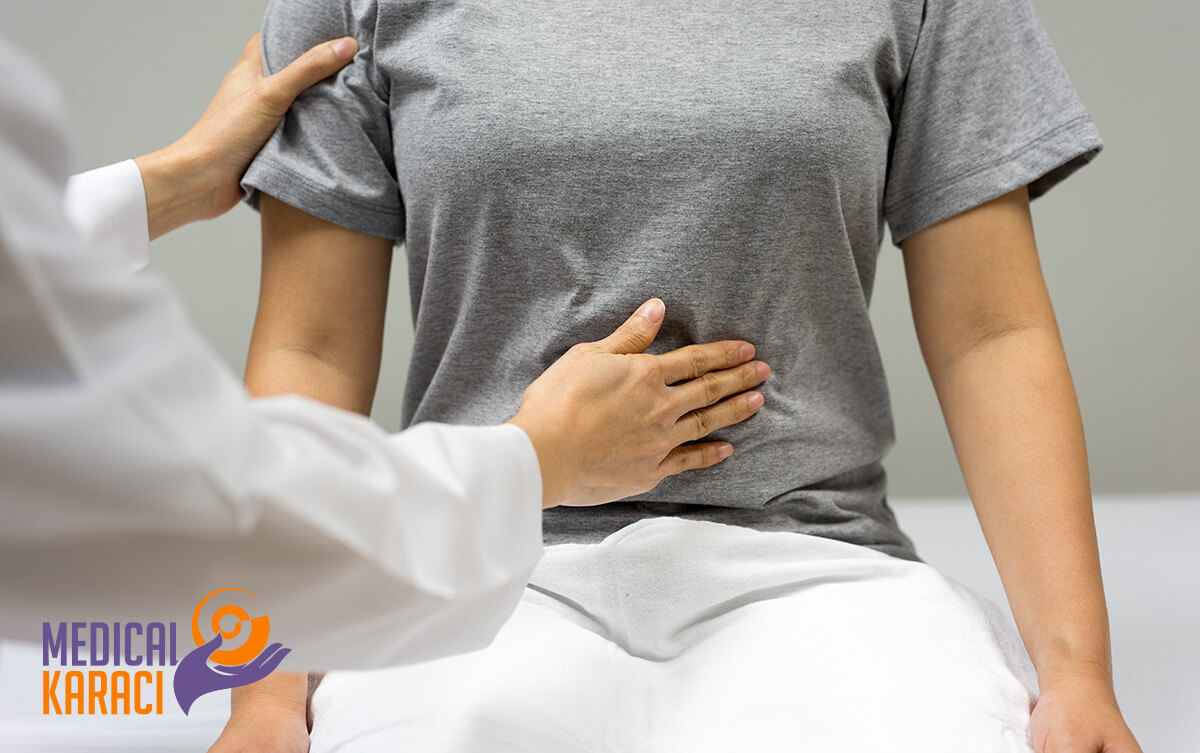
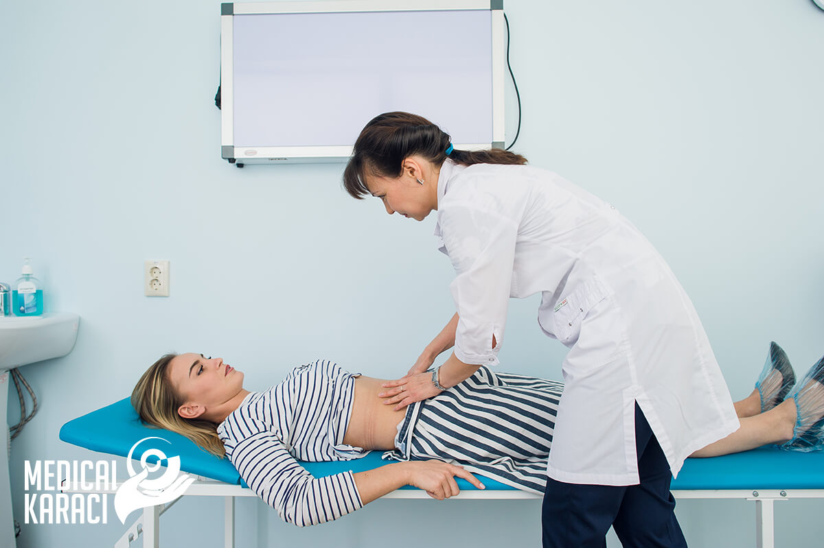
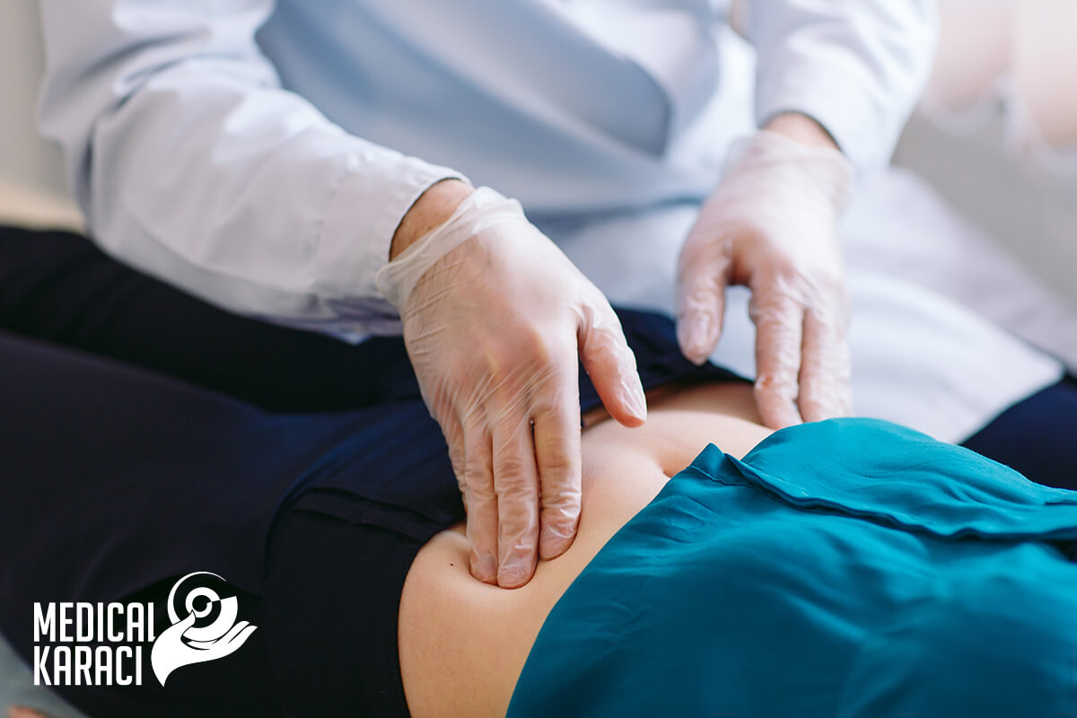
Gastroenterology is a branch of medicine that deals with the study, diagnosis, prevention and treatment of diseases of the human digestive system. It involves a detailed understanding of the normal functioning (physiology) of the gastrointestinal organs, including the movement of food through the stomach and intestines (motility), the digestion and absorption of nutrients in the body, the removal of waste from the system, and the function of the liver as a digestive organ.
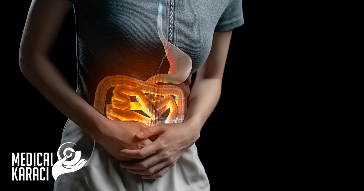
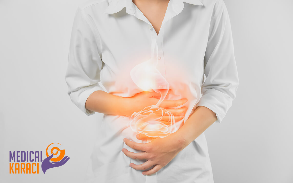
The organs of the digestive system are: tongue, salivary glands, epiglottis, pharynx, esophagus, stomach, liver, gallbladder, pancreas, small intestine, colon, rectum, anus.
Gastroenterologists can treat any part of this system.
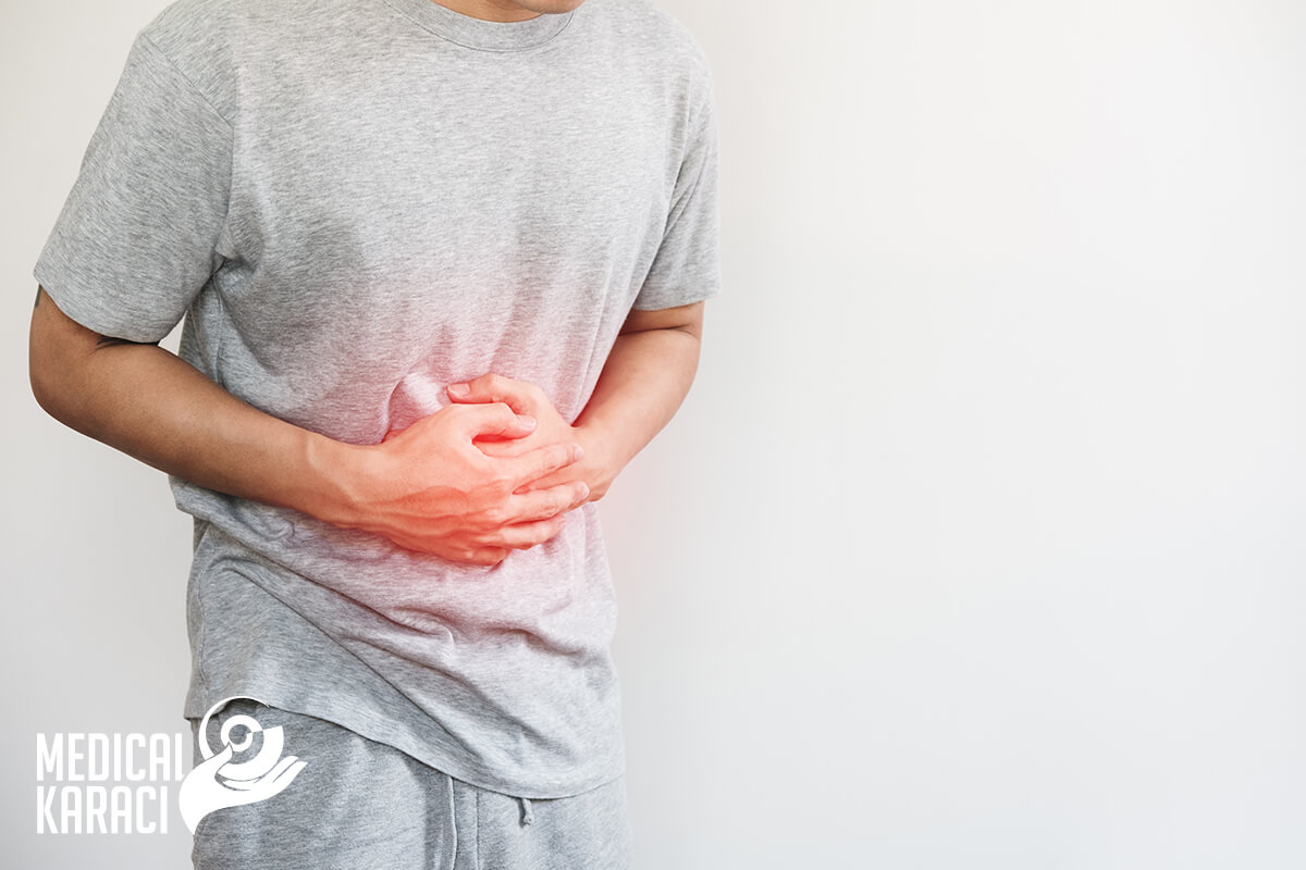
Some of the most typical diseases of the gastrointestinal tract are:
Ulcer of the stomach and duodenum
Gastric ulcers are painful sores in the lining of the stomach. Stomach ulcers occur when the thick layer of mucus that protects the stomach from digestive juices is reduced. This allows digestive acids to eat away at the tissues that line the stomach, causing an ulcer. Stomach ulcers can be easily healed, but can become severe without proper treatment.
Gastric ulcers are almost always caused by:
- infection with the bacterium Helicobacter pylori
- long-term use of non-steroidal anti-inflammatory drugs, such as aspirin, ibuprofen or naproxen
- A rare condition known as Zollinger-Ellison syndrome can cause stomach and intestinal ulcers by increasing acid production in the body. This syndrome is thought to cause less than 1% of all peptic ulcers.
Symptoms of gastric ulcer:
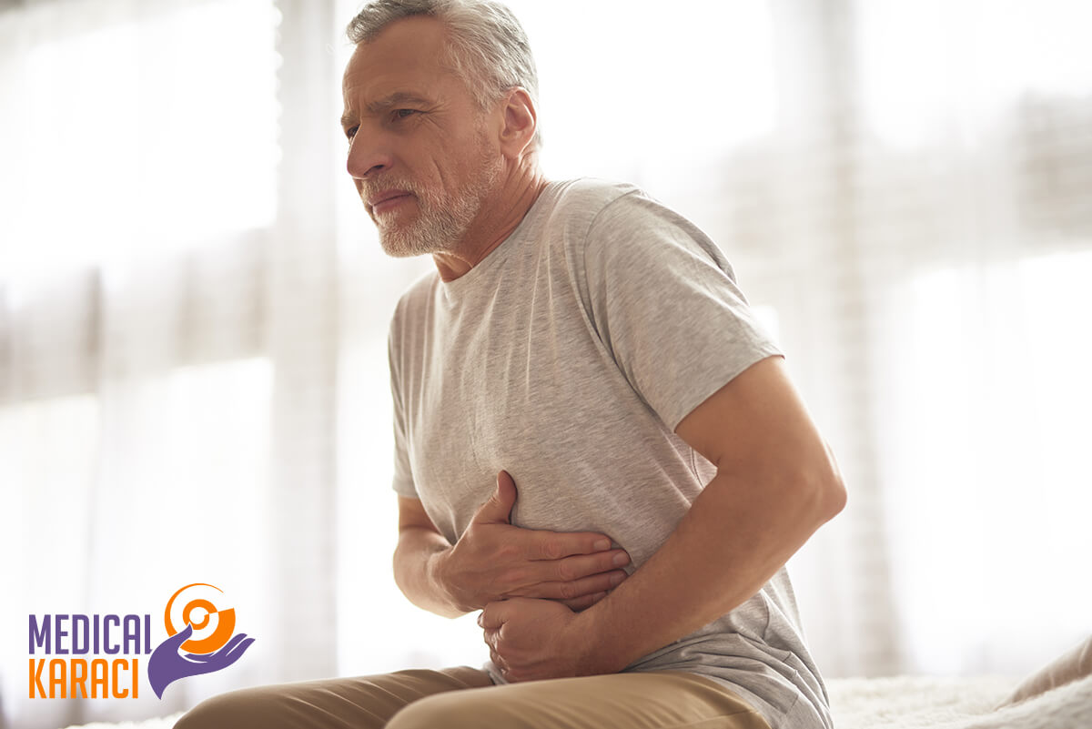
A number of symptoms are associated with stomach ulcers. The severity of symptoms depends on the severity of the ulcer. The most common symptom is a burning sensation or pain in the middle of the abdomen between the chest and the belly. The pain is usually more intense when the stomach is empty and can last from a few minutes to several hours.
Other common signs and symptoms of an ulcer include:
- dull pain in the stomach
- отслабване
- nausea or vomiting
- bloating
- belching or acid reflux
- heartburn, which is a burning sensation in the chest
- pain that may improve with eating, drinking or taking antacids
- anaemia, symptoms of which may include tiredness, breathlessness or paler skin
- dark, tarry stools
- vomiting that is bloody or looks like coffee grounds
Consult a gastroenterologist if you have symptoms of stomach ulcers. Although the discomfort may be mild, the ulcer can get worse if left untreated. Bleeding ulcers can become life-threatening.
Gastroesophageal reflux disease

Gastroesophageal reflux disease (GERD) is a digestive disorder that occurs when acidic gastric juices or food and fluids back up from the stomach into the esophagus. It usually occurs as a result of weakening of the ring of muscle at the bottom of the esophagus. GERD affects people of all ages, from babies to the elderly The disease causes symptoms such as heartburn and an unpleasant taste in the back of the mouth.
GERD can often be controlled with medication, but sometimes surgery may be needed to correct the problem.
Symptoms of GERD:
- heartburn - an uncomfortable burning sensation in the chest that often occurs after eating
- acid reflux - stomach acid goes back into the mouth and causes an unpleasant sour taste
- esophagitis - inflamed esophagus
- bad breath
- swelling and burping
- Nausea
- pain on swallowing and/or difficulty swallowing
Treatment of gastroesophageal reflux disease:
- dietary changes - this includes eating smaller amounts but more frequent meals, avoiding any foods or drinks that trigger symptoms, maintaining a healthy weight.
- over-the-counter medicines - antacid or alginate
- stronger prescription drugs - including proton pump inhibitors (PPIs) and H2-receptor antagonists (H2RAs)
You may only need to take medication when you have symptoms, although you may need long-term treatment if the problem gets worse.
Surgery to stop stomach acid leaking into the oesophagus may be recommended if medicines are not helping or you do not want to take them for a long time.
Stromal tumors of the gastrointestinal tract

Gastrointestinal stromal tumors are soft tissue sarcomas that can be localized in any part of the digestive system. Their most common sites are the stomach and small intestine. Gastrointestinal tumors begin in specialized nerve cells located in the walls of the digestive system. The exact cause of gastrointestinal stromal tumors is unknown. However, 95% of patients with gastrointestinal tumors have a protein called Kit (CD 117) that has become abnormal, which then causes normal cells to grow faster and become cancerous.
More than half of gastrointestinal stromal tumors begin in the stomach. Most of the rest start in the small intestine, but they can start anywhere in the digestive tract. A small number start outside the GI tract in nearby areas such as the omentum (the apron-like layer of fatty tissue that hangs over the organs in the abdomen) or the peritoneum (the thin lining over the organs and walls inside the abdomen).
It seems that some gastrointestinal stromal tumours are much more likely than others to grow into other areas or spread to other parts of the body. Doctors look at certain factors to see if a stromal tumor is likely to grow and spread quickly, such as:
- The size of the tumour
- Where it is found in the gastrointestinal tract
- How fast tumour cells divide
Other cancers of the gastrointestinal tract
Gastrointestinal stromal tumors are not the same as other, more common gastrointestinal cancers that develop from other types of cells.
Cancers can occur anywhere in the gastrointestinal tract - from the esophagus to the anus. Most cancers that begin in the gastrointestinal tract, including most esophageal cancers, stomach cancers, and colorectal cancers, begin in the cells of the glands that cover almost the entire gastrointestinal tract. Cancers that develop in these cells are called adenocarcinomas.
Cancers can also start in the squamous cells-the flat cells that line certain parts of the gastrointestinal tract, such as the upper esophagus and the end of the anus. Cancerous growths starting in these cells are called squamous cell carcinomas.
The gastrointestinal tract also has neuroendocrine cells. These cells have some features in common with nerve cells, but have other features in common with hormone-producing (endocrine) cells. Cancers that develop from these cells are called neuroendocrine tumors. These cancers are rarely found in the gastrointestinal tract. Carcinoid tumors are an example of a neuroendocrine tumor found in the gastrointestinal tract.
Other rare gastrointestinal cancers include various types of soft tissue sarcomas, such as:
- Leiomyosarcoma: cancer of the smooth muscle
- Angiosarcomas: cancer of blood vessel cells
- Malignant peripheral nerve sheath tumors: cancer of the cells that support and protect nerves
Gastrointestinal stromal tumors differ from these other types of gastrointestinal cancers. They start from different types of cells, need different types of treatment, and have a different prognosis (outlook). This is why doctors need to find out if a person with a tumor in the GI tract has a stromal tumor, some other type of cancer, or a noncancerous disease.
Crohn's disease
Crohn's disease is a type of inflammatory bowel disease. It causes inflammation of the digestive tract that can lead to abdominal pain, severe diarrhea, fatigue, weight loss and malnutrition. The inflammation caused by Crohn's disease can involve different areas of the digestive tract. This inflammation often spreads to the deeper layers of the intestine.
Symptoms:
In Crohn's disease, any part of the small or large intestine can be affected. In some people, the disease is limited to the colon only. The signs and symptoms of Crohn's disease can range from mild to severe. They usually develop gradually, but sometimes appear suddenly, without warning. There may also be periods of time when there are no signs or symptoms (remission).
When the disease is active, signs and symptoms may include:
- Diarrhoea
- High temperature
- Fatigue
- Abdominal pain and cramps
- Blood in your stool
- Sores in the mouth
- Decreased appetite and weight loss
- Pain near or around the anus due to inflammation from a fistula
People with severe Crohn's disease can also get:
- Inflammation of the skin, eyes and joints
- Inflammation of the liver or bile ducts
- Kidney stones
- Iron deficiency (anemia)
- Slow growth or sexual development in children
Reasons:
The exact cause of Crohn's disease remains unknown. Diet and stress used to be suspected, but doctors now know that these factors can aggravate but not cause Crohn's disease. Several factors, such as heredity and a malfunctioning immune system, likely play a role in its development.
- Immune system. It is possible for a virus or bacteria to trigger Crohn's disease. When the immune system tries to fight off the invading microorganism, an abnormal immune response causes it to also attack cells in the digestive tract.
- Heredity. Crohn's disease is more common in people who have family members with the disease, so genes may play a role in developing the disease. However, most people with Crohn's disease have no family history of the disease.
Complications:
Crohn's disease can lead to one or more of the following complications:
- Bowel obstruction. Crohn's disease can affect the entire thickness of the intestinal wall. Over time, parts of the intestine can narrow, which can block the flow of digestive contents. Surgery may be needed to remove the diseased part of the intestine.
- Scathing. Chronic inflammation can lead to open sores (ulcers) anywhere in the digestive tract, including the mouth and anus.
- Fistulas. Sometimes ulcers can spread entirely through the intestinal wall, creating a fistula - an abnormal connection between different parts of the body. Fistulas can develop between the intestine and the skin or between the intestine and another organ. Fistulas near or around the anal area (perianal) are the most common type. When fistulas develop in the abdomen, food can bypass areas of the intestine that are needed for absorption. Fistulas can form between the intestines, in the bladder or vagina, or through the skin, causing a continuous drainage of the intestinal contents to your skin. In some cases, the fistula can form an abscess, which can be life-threatening if left untreated.
- Anal fissure. This is a small tear in the tissue that damages the anus or the skin around the anus where infections can occur. It is often associated with painful bowel movements and can lead to a perianal fistula.
- Malnutrition. Diarrhoea, abdominal pain and cramps can make it difficult to eat or for the gut to absorb enough nutrients. Also, anemia often develops due to low iron or vitamin B-12 caused by the disease.
- Colon cancer. Crohn's disease increases the risk of colon cancer. General colon cancer screening guidelines for people without Crohn's disease require a colonoscopy every 10 years starting at age 50.
- Other health problems. Crohn's disease can cause problems in other parts of the body. These problems include anemia, skin diseases, osteoporosis, arthritis, and diseases of the gallbladder or liver.
- Risks of medication. Some Crohn's disease medications, which work by blocking immune system functions, are associated with a small risk of developing cancers such as lymphoma and skin cancer. They also increase the risk of infection. Corticosteroids may be associated with a risk of osteoporosis, bone fractures, cataracts, glaucoma, diabetes and high blood pressure, among other conditions.
- Blood clots. Crohn's disease increases the risk of blood clots forming in veins and arteries.
- Crohn's disease can be both painful and debilitating and can sometimes lead to life-threatening complications.
Although there is no known cure for Crohn's disease, therapies can significantly reduce its signs and symptoms and even lead to long-term remission and cure of inflammation. With treatment, many people with Crohn's disease are able to live well.
Ulcerative colitis
Ulcerative colitis is a disease that leads to inflammation and ulceration of the colon and rectum. The main symptoms of active disease are abdominal pain and diarrhea mixed with blood. Weight loss, fever and anemia may also occur.
The exact cause of ulcerative colitis is unknown, although it is thought to result from a problem with the immune system.
Autoimmune condition:
The immune system is the body's defence against infection. Many experts believe that ulcerative colitis is an autoimmune condition (when the immune system mistakenly attacks healthy tissue). The immune system normally fights infections by releasing white blood cells into the blood to destroy the cause of the infection. This causes swelling and redness (inflammation) of the body tissue in the infected area. In ulcerative colitis, a leading theory is that the immune system mistakes "friendly bacteria" in the colon that aid digestion as a harmful infection, leading to inflammation of the colon and rectum. Alternatively, some researchers believe that a viral or bacterial infection triggers the immune system, but for some reason it doesn't "turn off" once the infection is gone and continues to cause inflammation. It has also been suggested that it is not an infection and the immune system may simply be failing on its own, or that there is an imbalance between good and bad bacteria in the gut.
Genetics:
Inherited genes also appear to be a factor in the development of ulcerative colitis. Studies have found that more than 1 in 4 people with ulcerative colitis have a family history of the condition. Researchers have identified several genes that seem to make people more likely to develop ulcerative colitis. Many of these genes are thought to play a role in the immune system.
Environmental factors:
Where and how you live also seems to affect your chances of developing ulcerative colitis, suggesting that environmental factors are important. For example, the condition is more common in urban areas of northern Western Europe and America. Various environmental factors have been studied that may be linked to ulcerative colitis, including air pollution, medications and certain diets. Although no factors have yet been identified, countries with improved sanitation appear to have a larger population of people with this disease. This suggests that reduced exposure to bacteria may be an important factor.
Diagnosis of ulcerative colitis:
- Blood tests can show if there is anemia or inflammation.
- Stool samples can help rule out an infection or parasite in the colon. They can also show if there is hidden blood in the stool.
- Flexible sigmoidoscopy allows the lower part of the colon to be looked at. A flexible tube is inserted into the lower part of the colon. The tube has a small light and a camera at the end. A small instrument is used to take a piece of the lining of the lower colon. This is called a biopsy.
- Colonoscopy is the same process as flexible sigmoidoscopy, except that the entire colon is examined, not just the lower part.
- X-rays are less common to diagnose the disease, but may be assigned in special cases
Treatment of ulcerative colitis:
The treatment of ulcerative colitis has two main objectives. The first is to feel better and give the colon a chance to heal. The second is to prevent more exacerbations. You may need a combination of diet changes, medicines or surgery to achieve these goals.
Diet. Certain foods can worsen the symptoms of ulcerative colitis. A balanced diet with plenty of fibre, lean protein, fruit and vegetables should provide enough vitamins and nutrients.
Medication:
- Antibiotics - These fight infections and leave the colon to heal.
- Aminosalicylates - These medications have contain 5-aminosalicylic acid (5-ASA), which fights inflammation and helps control symptoms.
- Corticosteroids. If aminosalicylates do not work or the symptoms are severe, these anti-inflammatory drugs can be given as treatment for a short time.
- Immunomodulators - These help to stop the immune system's attack on the colon.
- Biological preparations. They are made of proteins instead of chemicals. They are used in people with severe ulcerative colitis.
- Surgery. If other treatments do not work or the condition is severe, surgery may be needed to remove the colon (colectomy) or colon and rectum (proctocolectomy). With a procolectomy, the small bowel can be connected to the anus and thus an ileostomy will be avoided.
Hepatitis
Hepatitis is an inflammatory disease of the liver. It is often caused by a viral infection, but there are other possible causes of hepatitis. These include autoimmune hepatitis and hepatitis that occurs as a secondary result of drugs, toxins and alcohol. Autoimmune hepatitis is a disease that occurs when the body produces antibodies against liver tissue.
Viral infections of the liver classified as hepatitis include hepatitis A, B, C, D and E. A different virus is responsible for each type of virally transmitted hepatitis.
- Hepatitis A - Hepatitis A is caused by infection with the hepatitis A virus (HAV). This type of hepatitis is most commonly transmitted by consuming food or water contaminated with stool from a person infected with hepatitis A.
- Hepatitis B - Hepatitis B is transmitted by contact with infected body fluids, such as blood, vaginal discharge or semen, containing the hepatitis B virus (HBV). Injecting drug use, having sex with an infected partner, or sharing razors with an infected person increase the risk of contracting hepatitis B.
- Hepatitis C - Hepatitis C is transmitted by direct contact with contaminated body fluids, usually through injection drug use and sexual contact.
- Hepatitis D - Also called delta hepatitis, hepatitis D is a serious liver disease caused by the hepatitis D virus (HDV). Infection occurs through direct contact with infected blood. Hepatitis D is a rare form of hepatitis that occurs only in association with hepatitis B infection. The hepatitis D virus cannot replicate without the presence of hepatitis B.
- Hepatitis E - Hepatitis E is a disease caused by the hepatitis E virus (HEV). Hepatitis E occurs mainly in areas with poor sanitation and usually results from ingestion of faecal matter that contaminates the water supply.
Causes of non-infectious hepatitis:
- Alcohol and other toxins
Excessive alcohol consumption can cause liver damage and inflammation. This is sometimes called alcoholic hepatitis. Alcohol directly damages liver cells. Over time, it can cause permanent damage and lead to liver failure and cirrhosis. Other toxic causes of hepatitis include overuse or overdose of medications and exposure to poisons.
- Autoimmune reaction
In some cases, the immune system takes the liver as a harmful object and begins to attack it. This causes inflammation and hinders liver function. It occurs more often in women than in men.
Common symptoms of hepatitis:
If you have infectious forms of hepatitis that are chronic, such as hepatitis B and C, you may have no symptoms at first. Symptoms may not appear until the damage affects liver function. Signs and symptoms of acute hepatitis appear quickly. They include:
- fatigue
- flu-like symptoms
- dark urine
- pale stools
- abdominal pain
- loss of appetite
- unexplained weight loss
- yellowing of the skin and eyes, which may be a sign of jaundice
Chronic hepatitis develops slowly, so these signs and symptoms may be too subtle to notice.
How is hepatitis diagnosed?
- Physical examination - abdominal pain or tenderness, enlarged liver, yellow skin and eyes
- Liver function tests - blood samples to determine how efficiently the liver is working. Abnormal results from these tests may be the first indication that there is a problem, especially if there are no signs of disease. High liver enzyme levels may indicate that the liver is stressed, damaged or not functioning properly.
- Ultrasound - Ultrasound of the abdomen may reveal: fluid in the abdomen, liver damage or enlargement, liver tumors, gallbladder abnormalities
- Liver biopsy - A liver biopsy is an invasive procedure that involves taking a sample of tissue from the liver. It can be done through the skin with a needle and does not require surgery. Ultrasound is usually used to guide the biopsy sample.
How is hepatitis treated?
Treatment options are determined by which type the hepatitis is and whether the infection is acute or chronic.
- Hepatitis A
Hepatitis A does not usually require treatment as it is a short-term disease. Bed rest may be recommended if the symptoms cause much discomfort. If vomiting or diarrhoea, hydration and nutritious feeding is needed. The hepatitis A vaccine is available to prevent this infection.
- Hepatitis B
Acute hepatitis B does not require specific treatment.
Chronic hepatitis B is treated with antiviral drugs. This form of treatment can be expensive because it must last for several months or years. Treatment for chronic hepatitis B also requires regular medical evaluations and monitoring to determine if the virus is responding to treatment. Hepatitis B can be prevented with vaccination.
- Hepatitis C
Antiviral drugs are used to treat acute and chronic forms of hepatitis C. People who develop chronic hepatitis C are usually treated with a combination of antiviral drug therapies. They may also need additional testing to determine the best form of treatment. People who develop cirrhosis or liver disease as a result of chronic hepatitis C may be candidates for a liver transplant. There is no vaccination against hepatitis C.
- Hepatitis D
There are currently no antiviral drugs available to treat hepatitis D. Hepatitis D can be prevented by vaccination against hepatitis B, since hepatitis B infection is necessary for the development of hepatitis D.
- Hepatitis E
No specific medical therapies are available for the treatment of hepatitis E. Because the infection is often acute, it usually resolves on its own. People with this type of infection are often advised to get adequate rest, drink plenty of fluids, get enough nutrients and avoid alcohol.
Autoimmune hepatitis:
Corticosteroids, such as prednisone or budesonide, are extremely important in the early treatment of autoimmune hepatitis. They are effective in about 80 percent of people with this condition. Azothioprine (Imuran), a drug that suppresses the immune system, is often included in treatment. It can be used with or without steroids. Other immunosuppressive drugs, such as mycophenolate (CellCept), tacrolimus (Prograf), and cyclosporine (Neoral) may also be used as alternatives to azathioprine for treatment.
Tips to prevent hepatitis:
- Hygiene - Practicing good hygiene is one of the key ways to avoid contracting hepatitis A and E.
- Hepatitis B, C and D, which is transmitted through contaminated blood, can be prevented by:
- not sharing drug needles
- not sharing razors
- not to use someone else's toothbrush
- not touching spilled blood
- Hepatitis B and C can also be contracted through sexual contact. Practicing safe sex by using condoms can help reduce the risk of infection.
- Vaccines - The use of vaccines is an important key to preventing hepatitis. Vaccinations are available to prevent the development of hepatitis A and B. Hepatitis C vaccines are currently being developed. Hepatitis E vaccination is available in China.
Complications of hepatitis:
Chronic hepatitis B or C can often lead to more serious health problems. Because the virus affects the liver, people with chronic hepatitis B or C are at risk for:
- chronic liver disease
- cirrhosis
- liver cancer
Cirrhosis of the liver
Cirrhosis is a severe chronic liver disease. Cirrhosis is most often caused by prolonged exposure to toxins such as alcohol or viral infections.
How does cirrhosis develop?
The liver is a very resilient organ and is usually able to regenerate damaged cells. Cirrhosis develops when factors that damage the liver,such as alcohol and chronic viral infections, are present for a long period of time. When this happens, the liver becomes injured and forms scarring/cyatrix. The liver scar cannot function properly and eventually this can lead to cirrhosis. Cirrhosis causes the liver to shrink and harden. This makes it difficult for nutrient-rich blood to flow into the liver from the portal vein. The portal vein carries blood from the digestive organs to the liver. The pressure in the portal vein rises when blood cannot pass into the liver. The end result is a serious condition called portal hypertension, in which the vein develops high blood pressure. This high-pressure system creates a backup that leads to esophageal varices such as varicose veins, which can then burst and bleed.
Common causes of cirrhosis:
- Alcohol
Cirrhosis can develop in women who drink more than two alcoholic drinks a day (including beer and wine) for many years. For men, drinking more than three drinks a day for years can put them at risk of cirrhosis. However, the amount is different for each person, and this does not mean that everyone who has ever had more than a few drinks will develop cirrhosis. Cirrhosis caused by alcohol is usually the result of drinking more than these amounts on a regular basis for 10 or 12 years.
Other causes of cirrhosis include:
- Hepatitis B can cause inflammation of the liver and damage that can lead to cirrhosis.
- Hepatitis D can also cause cirrhosis.
- Autoimmune hepatitis causes inflammation that can lead to cirrhosis.
- Damage to the bile ducts that function as bile drainage: an example of such a condition is primary biliary cholangitis.
- Disorders that affect the body's ability to handle iron and copper: for example, hemochromatosis and Wilson's disease.
- Drugs such as acetaminophen, some antibiotics and some antidepressants can lead to cirrhosis.
Symptoms of cirrhosis:
Symptoms of cirrhosis occur because the liver is unable to purify the blood, break down toxins, produce clotting proteins, and assist in the absorption of fats and fat-soluble vitamins. Often there are no symptoms until the disorder progresses. Some of the symptoms include:
- reduced appetite
- nosebleeds
- jaundice (yellow discoloration)
- small spider-shaped veins under the skin
- отслабване
- anorexia
- itchy skin
- Weakness
More serious symptoms include:
- confusion and difficulty thinking
- bloating (ascites)
- swelling of the feet (edema)
- Impotence
- gynecomastia (when men begin to develop breast tissue)
How is cirrhosis of the liver diagnosed?
The diagnosis of cirrhosis begins with a detailed history and physical examination. The history may reveal long-term alcohol abuse, exposure to hepatitis C, a family history of autoimmune disease, or other risk factors. Physical examination may reveal:
- pale skin
- yellow eyes (jaundice)
- red palms
- trembling of the hands
- enlarged liver or spleen
- small testicles
- excess breast tissue (in men)
- reduced alertness
Tests can reveal how damaged the liver is. Some of the tests used to assess cirrhosis are:
- complete blood count (to reveal anemia)
- blood tests for coagulation (to see how fast the blood is clotting)
- albumin (for testing protein produced in the liver)
- liver function tests
- alpha fetoprotein (liver cancer screening)
Additional tests that can evaluate the liver include:
- upper endoscopy (to see if there are esophageal varices)
- ultrasound scan of the liver
- MRI of the abdomen
- CT of the abdomen
- liver biopsy (the definitive test for cirrhosis)
Complications from cirrhosis:
If blood cannot pass through the liver, it creates a backup through other veins such as those in the esophagus. This backup is called esophageal varices. These veins are not built to handle the high pressure and begin to bulge from the extra blood flow.
Other complications of cirrhosis include:
- bruising (due to low platelet count and/or poor clotting)
- bleeding (due to reduced clotting of proteins)
- drug sensitivity (the liver processes drugs in the body)
- kidney failure
- liver cancer
- insulin resistance and type 2 diabetes
- hepatic encephalopathy (confusion due to the effects of blood toxins on the brain)
- gallstones (interference with bile flow can lead to hardening of the bile and stone formation)
- esophageal varices
- enlarged spleen (splenomegaly)
- oedema and ascites
Treatment of cirrhosis:
Treatment for cirrhosis varies depending on what caused it and how far the disorder has progressed. Some treatments include:
- beta-blockers or nitrates (for portal hypertension)
- quitting drinking (if cirrhosis is caused by alcohol)
- banding procedures (used to control bleeding from esophageal varices)
- intravenous antibiotics (to treat peritonitis that can occur with ascites)
- haemodialysis (to purify the blood of those with kidney failure)
- lactulose and low protein diet (for treatment of encephalopathy)
A liver transplant is a last resort when other treatments fail.
All patients should stop drinking alcohol. Medications, even over-the-counter ones, should not be taken without consulting a doctor.
Ventral hernia
A ventral hernia occurs along the vertical center of the abdominal wall. Symptoms include abdominal pain, especially when lifting or straining. Treatment is by surgery, including open, laparoscopic and robotic hernia reconstruction. A hernia occurs when there is a hole in the muscle of the abdominal wall, allowing a loop of bowel or abdominal tissue to push through the muscle layer. A ventral hernia is a hernia that occurs anywhere along the midline (vertical center) of the abdominal wall. There are three types of ventral hernia:
- Epigastric (stomach area) hernia: Occurs anywhere from just below the breastbone to the belly button. This type of hernia is seen in both men and women.
- Umbilical hernia: occurs in the navel area.
- Incisional hernia: develops at the site of a previous operation. Up to one-third of patients who undergo abdominal surgery will develop an incisional hernia at the site of the scar. This type of hernia can occur at any time, from months to years after abdominal surgery.
What are the causes and risk factors for developing a ventral hernia?
There are many reasons, including:
- Weakness at the incision site in previous abdominal surgery (which may be the result of an infection at the site of surgery).
- Congenital weakness in the abdominal wall area,.
- Weakness in the abdominal wall caused by conditions that strain the wall.
- Prostatism (enlargement of the prostate gland that causes straining to urinate in older men)
- Age (total loss of abdominal wall elasticity)
What are the signs and symptoms of a ventral hernia?
Some patients experience no discomfort in the early stages of ventral hernia formation. Often the first sign is a visible bulge under the skin in the abdomen or an area that is tender to the touch. The bulge may flatten when you lie down or squeeze it.
A ventral hernia causes an increasing level of pain when a person:
- Lifts heavy objects.
- When passing stool/urinating.
- Sitting or standing for long periods of time.
- Severe abdominal pain can occur if part of the bowel bulges through the abdominal wall and gets into the opening. If this happens, the trapped part of the intestine twists, loses its blood supply and begins to die. This is a medical emergency that requires immediate help.
How is a ventral hernia repaired?
Ventral hernias do not go away and do not get better on their own and require surgery to repair. In fact, without treatment, ventral hernias can become larger and worsen over time. Untreated hernias can become difficult to repair and can lead to serious complications, such as twisting part of the intestine. The goal of surgery for a ventral hernia is to repair the hole/defect in the abdominal wall so that the bowel and other abdominal tissue cannot bulge through the wall again. The surgery often restores the tone and shape of the abdominal wall, repairing the hole and returning the muscles to their normal position.
There are three main types of hernia repair surgery: open, laparoscopic and robotic:
- Open hernia repair surgery: an open incision is made in the abdomen where the hernia occurred, and the bowel or abdominal tissue is pushed back into place. A mesh material is placed to reduce recurrence. The skin is usually closed with dissolvable stitches and glue.
- Laparoscopic surgery: several small incisions are made where the hernia occurred. A laparoscope (a thin lighted tube with a camera on the tip) is inserted through one of the holes to help guide the surgery. Surgical mesh material may be inserted to strengthen the weakened area in the abdominal wall. Advantages of this approach compared with open hernia repair include less risk of infection as smaller incisions are used.
- Robotic hernia repair: like laparoscopic surgery, robotic surgery uses a laparoscope and is performed in the same way (small incisions, small camera). Robotic surgery differs from laparoscopic surgery because the surgeon is seated at a console in the operating room and handles the surgical instruments from the console. The advantages of robotic hernia surgery are that the patient has small scars rather than one large incision and may have less pain after this surgery compared to open surgery.
Inguinal hernia
An inguinal hernia occurs when bowel or abdominal fat bulges through the lower abdominal wall into the inguinal or groin area.
There are 2 types of inguinal hernias:
- Indirect inguinal hernias: this type of hernia is caused by a congenital defect in the abdominal wall
- Direct inguinal hernias: This type of hernia usually occurs in adult men. They are most often caused by weakness in the abdominal wall muscles that develops over time, or due to straining or heavy lifting.
An inguinal hernia is usually not dangerous. However, it can be painful, especially when lifting, bending, straining, when passing or coughing.
What are the symptoms of an inguinal hernia?
An inguinal hernia can be painless or cause no symptoms, especially when it first appears. Symptoms that may develop include:
- A bulge on one or both sides of the groin that disappears in the supine position.
- Groin pain, especially when lifting, coughing or straining.
- A feeling of weakness, heaviness, or burning in the groin.
- Swollen scrotum
Do all inguinal hernias require surgery?
An inguinal hernia does not get better or go away on its own. If you are diagnosed with an inguinal hernia, your doctor will probably recommend surgery.
How is an inguinal hernia repaired?
Inguinal hernia repair is a common surgical procedure. Inguinal hernia surgery is also called herniorrhaphy or hernioplasty. There are 3 types of inguinal hernia repair: open surgery,minimally invasive or laparoscopic repair and robotic repair
What are the risks of inguinal hernia repair surgery?
The risks of inguinal hernia repair include:
- Infection
- Bleeding
- Pain that is not relieved by medication
- Long-term complications are rare, but may include nerve damage or hernia recurrence, requiring a second operation.
Hiatal hernia
What is a hiatal hernia?
A hiatal hernia occurs when the upper part of the stomach pushes through an opening in the diaphragm and into the chest cavity. The diaphragm is the thin muscular wall that separates the chest cavity from the abdomen. The opening in the diaphragm is where the esophagus and stomach connect.
Who is at risk of developing a hiatal hernia?
Hiatal hernia can develop in people of all ages and both sexes, although it is common in people over the age of 50. Hiatal hernia is more common in overweight people and smokers.
What are the symptoms of hiatal hernia?
Many people with hiatal hernia have no symptoms. Some people have the same symptoms as gastroesophageal reflux disease (GERD).
- Heartburn.
- Bitter or sour taste in the back of the throat.
- Swelling and belching.
- Discomfort or pain in the stomach or esophagus.
Although there appears to be a link between hiatal hernia and GERD, one condition does not cause the other. Many people have a hiatal hernia without having GERD, and others have GERD without having a hiatal hernia.
Another symptom of hiatal hernia is chest pain.
How is hiatal hernia diagnosed?
Several tests can be done to help diagnose a hiatal hernia. These include a barium ingestion test, endoscopy, esophageal manometry studies, pH test, and gastric emptying studies.
How is hiatal hernia treated?
Most hiatal hernias do not cause problems and rarely need treatment. However, because some patients with hiatal hernia have symptoms of GERD, treatment begins with methods used to treat GERD.
When is hiatal hernia surgery necessary?
If the part of the stomach entering the esophagus is squeezed so hard that the blood supply is cut off, surgery will have to be done. Surgery may also be needed for people with hiatal hernia who have severe, long-lasting (chronic) oesophageal reflux whose symptoms are not relieved by medical treatments. The goal of this surgery is to correct gastroesophageal reflux by creating a flap mechanism at the bottom of the esophagus.
How is hiatal hernia surgery performed?
Hiatal hernia repair surgery includes:
- Pulling the hiatal hernia back into the abdomen.
- Improving the valve at the bottom of the esophagus.
- Closure of the hole in the diaphragm muscle.
During surgery, the upper part of the stomach(called the fundus) is wrapped around the lower part of the esophagus. This creates a permanently tight sphincter (valve) so that stomach contents do not move back (reflux) into the esophagus. The operation can be open or laparoscopic.
Open fundoplication surgery involves a larger incision. This type of procedure may need to be done in some very severe cases and allows greater visibility during surgery. However, open surgeries require a longer recovery time in the hospital.
Laparoscopic surgery is performed through several small incisions instead of one large incision. The specific laparoscopic procedure used for hiatal hernia repair is called Nissen fundoplication. This procedure creates a permanent solution to the symptoms of a hiatal hernia. During the procedure, five or six small incisions are made in the abdomen. The laparoscope and other surgical instruments are inserted through the small incisions. The upper part of the stomach is wrapped around the esophagus and the sphincter is tightened during the surgery. Advantages of laparoscopic surgery over open surgery include:
- Smaller incisions.
- Less risk of infection.
- Less pain and scarring.
- Shorter recovery.
Haemorrhoids
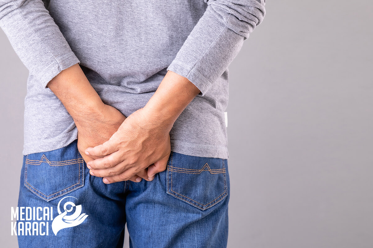
Hemorrhoids are swollen veins located around the anus or in the lower part of the rectum. About 50 percent of adults experience the symptoms of hemorrhoids by age 50.
Hemorrhoids can be both internal and external. Internal hemorrhoids develop in the anus or rectum. External hemorrhoids develop outside the anus. External hemorrhoids are the most common and troublesome. Hemorrhoids cause pain, severe itching, and difficulty sitting. Fortunately, they are treatable.
What are the symptoms of hemorrhoids?
Symptoms of hemorrhoids include:
- severe itching around the anus
- irritation and pain around the anus
- itchy or painful lump or swelling near the anus
- leakage of faeces
- painful bowel movements
- blood on the tissue after bowel movement
Although hemorrhoids are painful, they do not pose a threat to life and often disappear on their own without treatment.
What causes hemorrhoids?
Hemorrhoids occur when there is too much pressure on the veins around the anus. Possible factors include:
- tension during bowel movement
- complications of chronic constipation
- sitting for long periods of time, especially on the toilet
- family history of haemorrhoids
Risk factors:
- Hemorrhoids can be passed genetically from parent to child.
- Constant weight lifting, obesity or other constant strain on the body can increase the risk of hemorrhoids.
- Hemorrhoids can develop when straining during bowel movements whether you have diarrhea or constipation or sit too long on the toilet.
- Anal intercourse can also irritate hemorrhoids.
- You are also more likely to develop hemorrhoids during pregnancy. When the uterus enlarges, it squeezes the vein in the colon, causing it to bulge.
How are hemorrhoids diagnosed?
Visual examination of the anus may be sufficient to diagnose hemorrhoids. To confirm the diagnosis, a different examination can be done to check for abnormalities in the anus. This examination is known as a rectal examination. During this examination, the doctor inserts a finger into the rectum. Depending on the risk factors for gastrointestinal diseases, the doctor may order an additional test such as anoscopy, sigmoidoscopy or colonoscopy. Each of these tests involves using a small camera to diagnose abnormalities in the anus, rectum or colon.
Anoscopy examines the inside of the anus, sigmoidoscopy examines the last 40 centimeters of the colon, and colonoscopy examines the entire colon.
What are the treatment options for hemorrhoids?
If home treatments do not help with hemorrhoids, the doctor may recommend to do ligation with an elastic band. This procedure involves the doctor cutting off blood circulation to the hemorrhoid by placing an elastic band around it. This causes the hemorrhoid to lose circulation, forcing it to shrink. This procedure should only be performed by a medical professional.
If ligation with an elastic is not an option, the doctor may perform injection therapy or sclerotherapy. In this procedure, a chemical is injected directly into the blood vessel. This causes the hemorrhoid to decrease in size.
What are the complications associated with hemorrhoids?
Complications from hemorrhoids are rare, but can include:
- blood clots in the swollen vein
- Bleeding
- iron deficiency anemia caused by blood loss
Dyspepsia
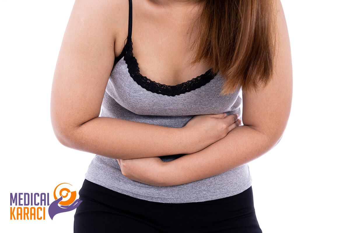
Dyspepsia, also known as indigestion, refers to discomfort or pain that occurs in the upper abdomen, often after eating or drinking. It is not a disease, but a symptom. Dyspepsia is a common problem that affects up to 30% of the population. Common symptoms include bloating, discomfort, feeling too full, nausea and gas. In most cases this occurs after eating or drinking. Lifestyle changes can often help.
Other causes include medical conditions such as gastroesophageal reflux disease (GERD) and the use of certain medications.
Symptoms:
- pain associated with the digestive system
- burning sensation in the digestive tract
- feeling full after eating
- feeling full too quickly during a meal
- One may also experience swelling and nausea.
- A person can have symptoms even if he has not eaten a large amount.
Treatment:
Treatment of dyspepsia depends on the cause and severity. Often treating the underlying condition or changing a person's medication will reduce the dyspepsia.
For mild and rare symptoms, lifestyle changes can help. These include:
- avoiding or limiting intake of trigger foods, such as fried foods, chocolate, onions and garlic
- drinking water instead of soda
- limit caffeine and alcohol intake
- eating smaller amounts more often
- slow eating
- maintaining a moderate weight
- avoiding tight-fitting clothing
- without eating 3 hours or more before bedtime
- raising the head of the bed
- avoiding or quitting smoking
With severe or frequent symptoms, the doctor may recommend medication. Different medications and treatments are available, depending on the cause of the dyspepsia.
Some of the most common interventions for diseases of the gastrointestinal tract are:
Laparoscopic appendectomy
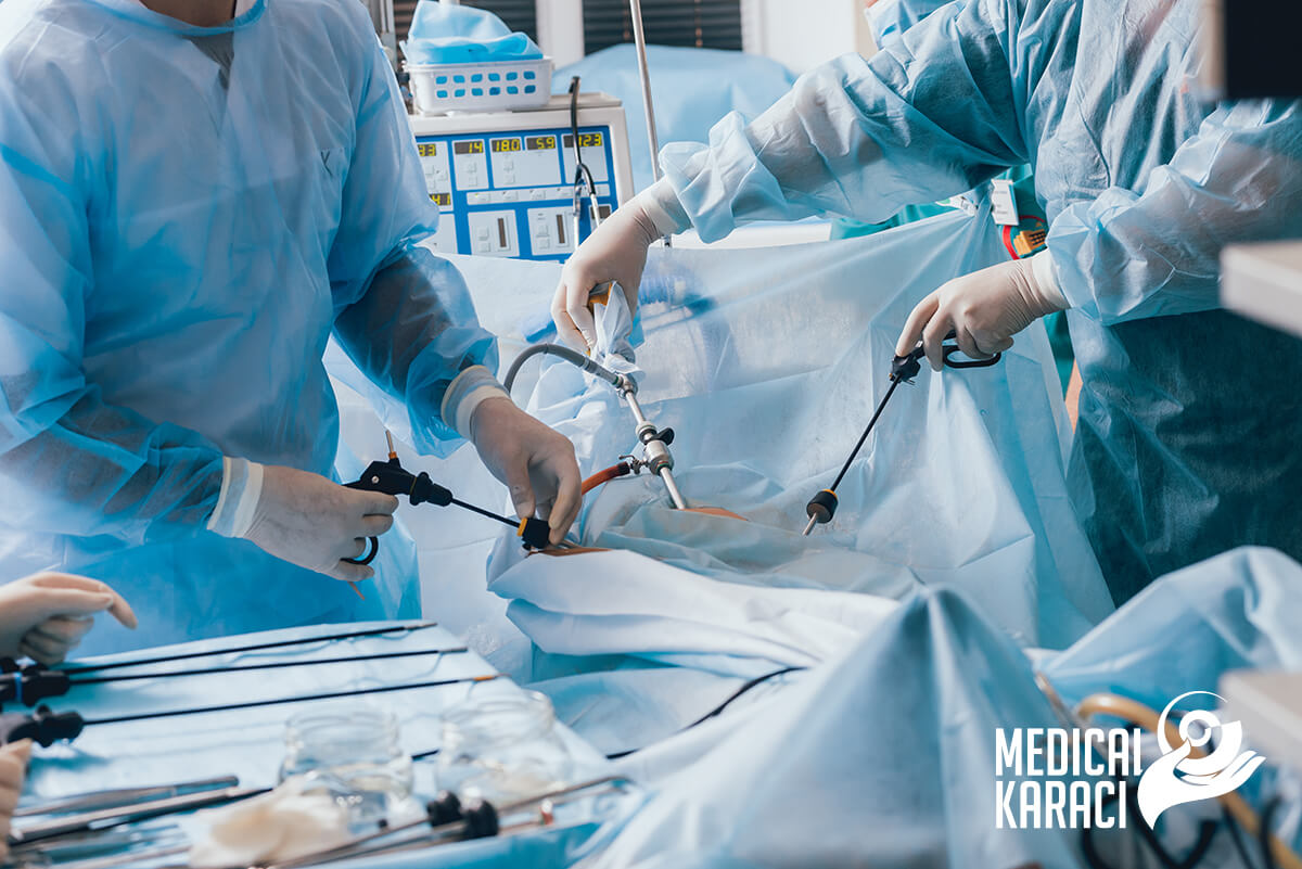
What is an appendectomy?
This is a type of surgery to remove the appendix. Removing the appendix cures appendicitis. If the appendix is not treated, it can burst and cause very serious illness or even death.
Appendectomy is a common operation. One way to remove the appendix is an open appendectomy. Laparoscopic appendectomy removes the appendix using small incisions.
Laparoscopic cholecystectomy
What is laparoscopic cholecystectomy?
Laparoscopic cholecystectomy is surgery to remove the gallbladder. The operation takes about1-2 hours. Laparoscopic cholecystectomy helps people with gallstones that cause pain and infection. Gallstones are crystals that form in the gallbladder. They can block the flow of bile from the gallbladder into the digestive system. This obstruction causes cholecystitis (inflammation of the gallbladder). Gallstones can also move to other parts of the body and cause problems.
Symptoms of gallstones include:
- A feeling of bloatedness.
- High temperature.
- Jaundice.
- Nausea.
- Pain in the right side of the abdomen that may reach the back or shoulder.
Gastroscopy
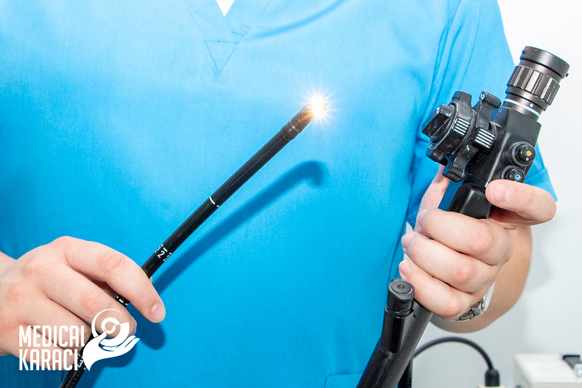
What is gastroscopy?
Gastroscopy is an examination of the upper digestive tract (esophagus, stomach and duodenum) using an endoscope - a long, thin, flexible tube containing a camera and light to see the lining of these organs.
Why is a gastroscopy done?
Gastroscopy, also known as upper gastrointestinal endoscopy or simply upper endoscopy, is usually done to examine the cause of symptoms such as heartburn, abdominal pain, difficulty swallowing, vomiting or bleeding from the digestive tract, as well as to make or confirm a diagnosis.
Sometimes conditions can also be treated during gastroscopy - for example:
- polyps can be removed;
- Varicose veins (boils) in the esophagus can be treated to stop and prevent bleeding;
- narrowed esophagus can be dilated
- foreign objects (such as things swallowed by children) can be removed.
The procedure takes about 15 to 30 minutes. You can usually go home after about 2 hours.
What are the risks of gastroscopy?
After the procedure, there may be a slight sore throat.Air may get into the stomach, causing swelling.
There is a small chance of bleeding after the procedure, especially if a biopsy was taken or treatment was performed. Any bleeding is usually minor.
Very few people experience serious side effects from gastroscopy. Very rarely, the lining of the stomach, oesophagus or duodenum (the first part of the small intestine) may be torn and in such cases hospitalisation is necessary. Surgery may be required to repair the perforation.
Colonoscopy
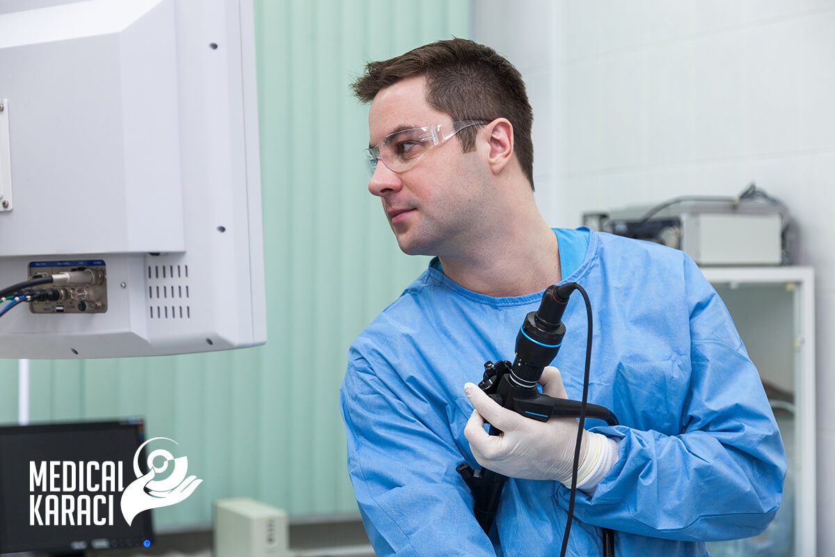
What is a colonoscopy?
A colonoscopy is a procedure in which the doctor uses a colonoscope to look inside the rectum and colon. A colonoscopy can show irritated and swollen tissue, ulcers, polyps, and colon tumors.
A colonoscopy can help the doctor find the cause of symptoms, such as:
- bleeding from the anus
- changes in bowel activity, such as diarrhea
- abdominal pain
- unexplained weight loss
Doctors also use colonoscopy as a screening tool for colon polyps and cancer. Screening can detect diseases at an early stage when there is a better chance of curing the disease.
For the smooth conduct of the examination, preliminary preparation is necessary - cleaning the colon (emptying it of feces). The whole procedure of preparation for colonoscopy is explained by the doctor at the preliminary consultation.
Proper preparation is very important. A poorly cleaned bowel can be the cause of missed small polyps or tumors. Inspection is difficult in the presence of foul water and food debris.
A colonoscopy usually takes 30 to 60 minutes.
During the procedure, the doctor may remove polyps and send them to a laboratory for testing. Colon polyps are common in adults and in most cases are harmless. However, most cases of colon cancer begin as a polyp, so early removal of polyps helps prevent cancer.
If the doctor finds abnormal tissue a biopsy may be performed. If the doctor has removed polyps or performed a biopsy, there may be slight bleeding from the anus. This bleeding is normal. The pathologist will examine the biopsy tissue, and the results take several days or longer.
What are the risks of colonoscopy?
Risks of colonoscopy include:
- Bleeding
- perforation of the colon
- reaction from the anaesthetic, including breathing or heart problems
- severe abdominal pain
- death, although this risk is rare
Bleeding and perforation are the most common complications of colonoscopy. Most cases of bleeding occur in patients in whom polyps have been removed.
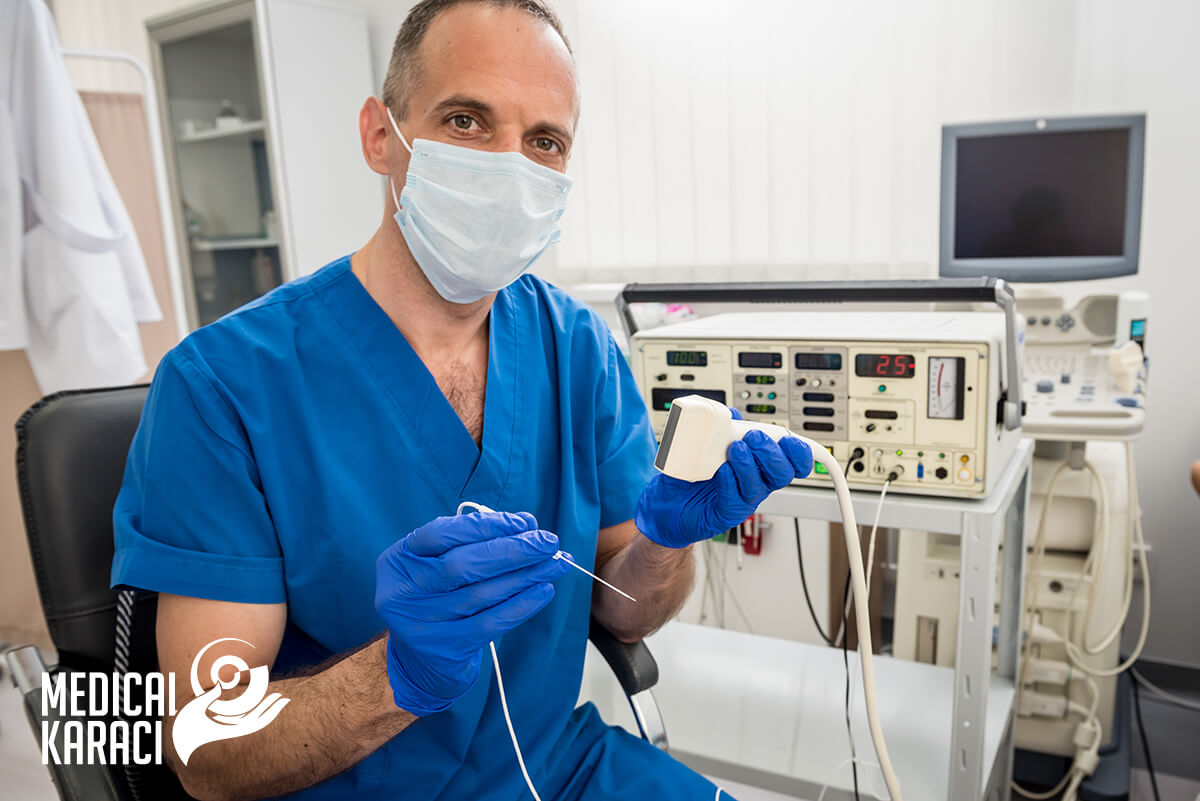
Ultrasound diagnostics

ltrasound diagnostics is used to check the main organs in the abdominal cavity. These organs include the gallbladder, kidneys, liver, pancreas and spleen.
If the specialist suspects that you have any of these conditions, he or she may do an abdominal ultrasound:
- blood clot
- enlarged organ (such as liver, spleen or kidney)
- fluid in the abdominal cavity
- gallstone
- hernia
- Pancreatitis
- kidney obstruction or cancer
- kidney stone
- liver cancer
- appendicitis
- tumour formations
Abdominal ultrasonography can also be used to help during certain procedures. For example:
- During an abdominal biopsy to see where to place the needle to remove a small sample of tissue.
- Ultrasound can help drain fluid from a cyst or abscess.
- Ultrasound can be used to examine the blood flow inside the abdomen.
Radiofrequency thermal ablation
Radiofrequency neurotomy uses heat generated by radio waves to target certain nerves and temporarily shut down their ability to send pain signals. The procedure is also known as radiofrequency ablation. Needles inserted through the skin near the painful area deliver the radio waves to the targeted nerves.
Radiofrequency ablation is most commonly used to treat Barrett's esophagus, a condition that can often result from heartburn or gastroesophageal reflux disease, and gastric antral vascular ectasia, a condition of abnormal blood vessels in the stomach that causes anemia.
Helicobacter pylori test

Helicobacter pylori is a gram-negative bacterium that is found in the stomach and duodenum. It causes peptic gastritis, ulcer, duodenitis. It is suspected to be one of the factors in causing stomach cancer.
It is estimated that over 50% of the world's population is a carrier of this bacterium, mostly in developing countries. In 80% of cases, there is no symptomatology. In the infection itself, there may be nausea, vomiting, lack of appetite, but there may be no symptoms. The bacterium may develop for years without the patient suspecting it.
The study is recommended for:
- Prevention.
- The presence of discomfort in the gastric region.
- Diagnosed disease in the gastric region without an identified causative agent.
For more information, you can call +359895770869.