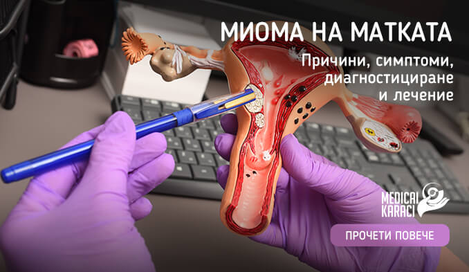
Uterine fibroids (also known as myomas) are benign neoplasms that develop in the muscular wall of the uterus. Approximately 20% of women suffer from uterine fibroids at reproductive age (especially women over 30). The size of fibroids can be microscopic and slowgrowing, which is why many women go undiagnosed in the beginning despite their complaints. Fibroids can, however, grow rapidly and even fill the entire uterine cavity. Multiple uterine fibroids of different shapes are also common.
CAUSES OF UTERINE FIBROIDS
The specific cause of myomatous uterus is not known, but nevertheless one of the main causes is thought to be related to genetic predisposition. Another factor playing a role in the development of fibroids is the hormonal activity of the ovaries, and in particular, the balance between estrogen and progesterone. These two hormones guide the formation and development of the uterine lining during each menstrual cycle and in preparation for becoming pregnant.
For this, myomas most often appear and grow during pregnancy, and decrease in size (even sometimes disappear) after the onset of menopause, when hormone levels are lower.
Where can the myoma be located ?
The location of uterine fibroids is of great importance as this determines whether they can cause infertility by altering sperm transport and/or the embryo implantation process.
- Intramural - the myoma is located in the muscular wall of the uterus, which affects fertility if the size is over 4cm
- Submucosal - the myoma is on the surface of the uterine mucosa. In these cases, implantation of the embryo in the uterine mucosa is difficult
- Subserosal - the myoma is in the outer lining of the uterus. This position is the most favorable, since the chance to affect fertility is very small
WHAT ARE THE MAIN SYMPTOMS OF UTERINE FIBROIDS?
About a quarter of women with fibroids have no symptoms. The majority of women exhibit symptoms such as:
- Cramps in the lower abdomen
- Excessive menstrual bleeding
- Extremely long or painful menstrual periods
- Bleeding outside menstruation
- Difficult or painful urination
- Infertility
- Recurrent miscarriages
It is important to emphasize that the symptoms and their severity are related to the size, location and number of fibroids.
In addition to difficulties getting pregnant, myoma poses other reproductive risks:
- Increased risk of miscarriage
- Slowing or stopping fetal growth
- Premature birth due to insufficient uterine space
- Obstruction of the cervical canal
- Postpartum haemorrhage
HOW ARE UTERINE FIBROIDS DIAGNOSED?
- Laboratory tests are mostly recommended in cases of abnormal uterine bleeding to point towards finding the cause. A complete blood count is done (to detect possible anemia from the blood loss), sex hormones, thyroid hormones, specialized tests for bleeding disorders.
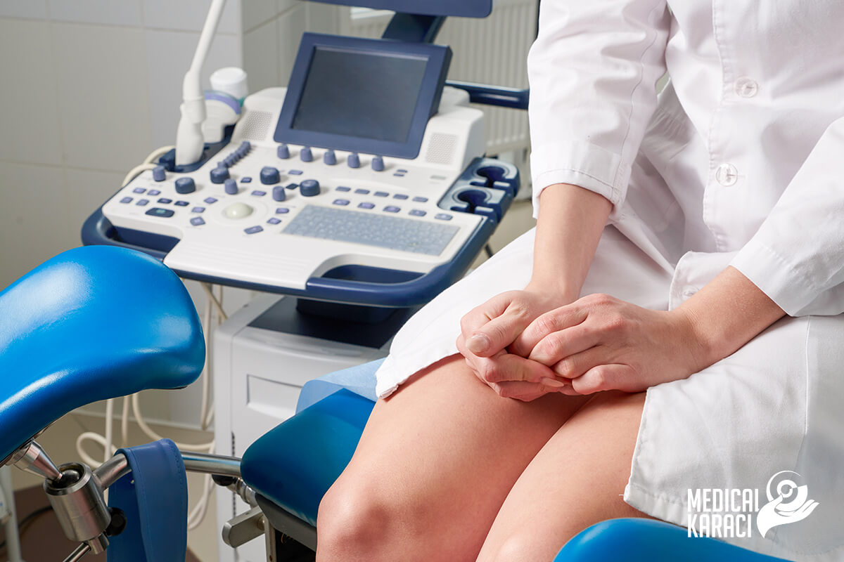
- Uterine fibroids are most commonly detected during ultrasonographic examination, with an accuracy of approximately 95% transabdominally and 100% when performed transvaginally.
- In case the patient shares complaints and the traditional ultrasound does not provide enough information, specialists may appoint other imaging studies such as:
- Hysterosonography (sonogram) - through sterile saline, the uterine cavity is dilated, which facilitates the imaging of the uterine mucosa, as well as the identification of submucosal myomas.
- Hysterosalpingography (color photo) - a commonly used diagnostic method for infertility. The injection of dye into the uterus allows the uterine cavity and fallopian tubes to be highlighted on the X-ray image. By means of the colour photograph, in addition to myomas, blockages of the fallopian tubes are also detected.
- Hysteroscopy - After injecting saline into the uterus, a small lighted telescope (hysteroscope) is passed through the cervix. In this way, a detailed examination of the uterine wall and fallopian tubes is achieved.
- Magnetic resonance imaging - thus giving more detailed information about the size and location of the myoma, as well as to make a differential diagnosis with different types of malignant formations. MRI is the preferred diagnostic method in women with larger uterine size, as well as in women approaching menopause.
TREATMENT
There are many cases in which small uterine fibroids do not pose a risk to a woman's health and doctors only recommend regular monitoring.
As for patients with myomatous uterus wishing to become pregnant, the case should be thoroughly analyzed and treated adequately. The treatment of myoma depends on the age of the woman, and especially on the size and location of the uterus.
As mentioned, subserosal myomas rarely affect fertility, while myomas in the lining or in the muscle tissue of the uterus require treatment (especially when they are larger than 5 cm and the woman is over 35 years of age). They can be an obstacle to sperm transport and subsequent implantation. When myomas affect the endometrial cavity, it is advisable to perform removal of the myoma (myomectomy), after which it is most appropriate to proceed to in vitro fertilization.
Conservative treatment:
- Oral contraceptive medications are used, which rather have a symptomatic treatment (reduce extra-menstrual bleeding or dysmenorrhea, and balance the thickness of the mucous membrane). By taking gonadotropin-regulating hormone agonists, the action of estrogen and progesterone on the myoma is blocked, achieving a state of temporary postmenopause. Thus, the myoma shrinks and the anaemic state improves.
- Another method of conservative treatment is the use of intrauterine devices with levonorgestrel - the well-known spiral - Mirena, for example. It is mainly used in women after the age of 35, regulating bleeding, but the size of the myoma often remains unchanged.
- An increasingly common method of treatment is the use of progesterone receptor modulators (SPRM).
In the late 1990s, the first non-surgical approach to myoma treatment or so-called radiologically guided arterial myoma embolization was introduced. - Nowadays, high-frequency ultrasound (HIFU) treatment is used as a current non-invasive method. Focused ultrasound waves are used to create zones of thermal coagulation in the myomas, which in turn leads to necrosis and shrinkage of the myoma.
- Fortunately, today there are also minimally invasive techniques with much greater success, among which is laser laparoscopy. The biggest benefits for women planning to be mothers are: no bleeding, risk of adhesions reduced to MINIMUM, and categorical preservation of ovarian reserve. Patients regain their normal rhythm of life much faster as the abdominal wall is not traumatized.
За повече информация, ние, Медикъл Караджъ сме на ваше разположение: 0878500730
Also keep an eye on our constantly updated Facebook content.