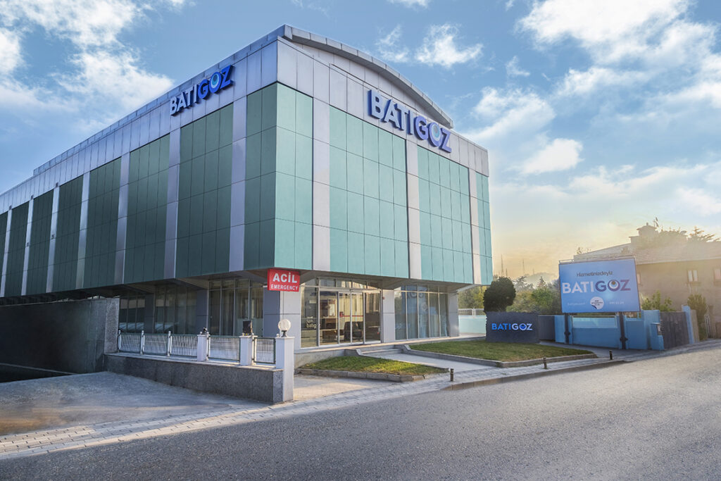
Batıgöz & Westeye Health Group is a renowned medical institution specializing in eye care. The company was established in 2004 by Op. Dr. Mehmet Soyler, Chairman of the Board and has successfully grown under his leadership, now covering 6 hospitals and 4 control centers in 6 different cities in 4 different countries. The company's success is based on two pillars:
- Understanding the patient .
- Technology and know-how.
The company is well positioned to treat both domestic and international patients,
And it has a steady growth path planned to double by 2020.
Our vision is to be the leading eye care provider in Turkey and abroad by establishing a network of control and diagnostic centers in Turkey and abroad by 2020, keeping optimal patient and employee satisfaction as our main goal.


Our mission is to understand the needs of our patients, to provide a wide range of high quality eye care treatments to every patient within our network, backed by state-of-the-art technology and research.
- TS EN ISO 9001 Quality Management System
- OHSAS 18001 Occupational Health and Safety Management System
- TS EN ISO 14001 Environmental Management System
- TS ISO 10002 Customer Satisfaction Management System
Why choose us?
Technological trends
The latest diagnostic and therapy options are available at all our centres and hospitals and we are continually investing in this.
Make sure we continue to follow the highest possible standards. Many technologies such as 3D imaging, Femtosecond Laser, Excimer Laser, Intralase IFS, ReLEx Smile and Argon Laser have been successfully deployed at Batıgöz and Westeye Healthcare Group.
Experience and education
Our doctors and nurses are at your service.
Educated in the best and reputable medical faculties in Turkey and abroad.
Our senior management, as well as our Chief Medical Officer, carefully plans continuing medical education and provides opportunities for medical staff to train and attend conferences in Turkey and abroad. All know-how is carefully balanced in a friendly working environment and constant monitoring of patients' rights.
In our hospitals in Izmir, Istanbul as well as Bucharest we offer successful treatment to international patients. We stand by our service and the medical quality offered throughout the group. We have multilingual qualified support staff as well as documentation processing. We ensure a smooth service and stay for all international patients.
Powerful medical infrastructure
All operating and laser rooms as well as examination rooms are fully equipped with special ventilation and air freshening systems.
A refreshment system that keeps the environment at the highest level of hygiene. All medical equipment and patient data across the world and the entire group is stored in a centralised location where we can analyse all information about our patients' therapy, testing and follow-up.
Health tourism
The Batıgöz and Westeye hospitals in Istanbul, İzmir and Bucharest are well equipped to serve international patients for any type of treatment required. Over the years, we have gradually built on this experience and offer a wide range of such professional services at our hospitals through our medical and concierge services as such:
- 24/7 operational and multilingual call centre
- English-speaking medical staff
- Professional hosts who can help in Russian, German, Dutch, English, French, Spanish and Arabic
- Full assistance and guidance from flight attendants during examinations as well as during operations and post-operations
- All documents and medical reports in Russian, German, Dutch, English, French, Spanish and Arabic
- Insurance intermediary services
- Transfer - airport - hotel - hospital transfer
- Professional tourism team ready to help you with any kind of tours and accommodation throughout Turkey or Romania

Assoc. Prof. Dr. Sevil Ara Yailali
Epiretinal membrane
The epiretinal membrane (ERM) is a thin, translucent tissue that appears on the inner surface of the central retina. The central retina, called the macula, is responsible for seeing fine details in reading, facial recognition, and driving.
In most cases, ERM is caused by an abnormality in age-related changes in aging. ERM can also occur in patients with diabetes, retinal blood vessel obstruction, uveitis and previous retinal surgery.
In the majority of cases, ERM causes no complaints and is detected incidentally on ophthalmological examination. However, thickening and shrinking of the membrane over time causes wrinkling of the macula. This leads to blurring of vision or revealing of images.
A diagnosis of ERM is made with a retinal examination and a specific retinal scan.
If ERM affects vision, the only way to treat it is surgical removal of the membrane. During surgery, the vitreous and ERM are removed through a procedure called vitrectomy.
Assoc. Prof. Dr. Sevil Ara Yailali
Figure 1:
In the scanned image on the left, the macula is deformed by ERM (left, white line on the retinal surface, arrow); the macula in the other eye of the same patient is normal (right).
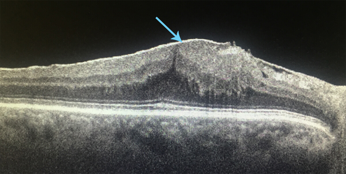
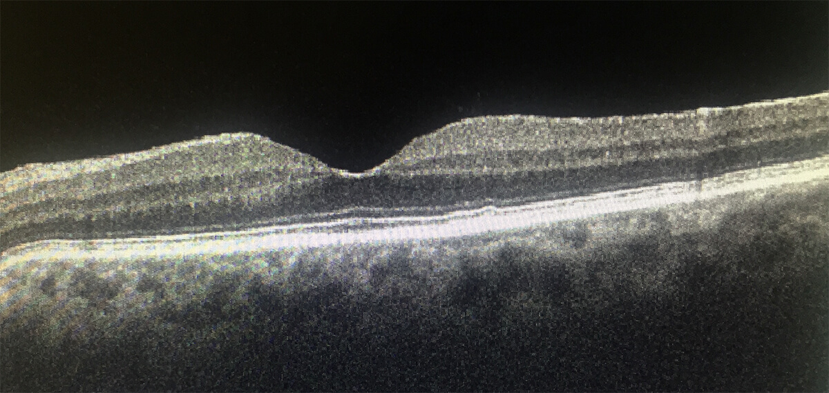
Figure 1
Figure 2:
A patient with ERM may see the lines distorted.
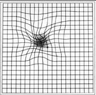
Figure 2
Macular hole
The macula is the central part of the retina that is responsible for seeing fine details in reading, facial recognition and driving.
A macular hole is a hole in the central retina. This condition often affects people aged 60 - 80 and is more common in women than men. The cause of macular holes has not been fully established. However, the abnormality of age-related detachment of the vitreous gel from the retina is thought to be a possible risk factor.
Patients may complain of blurred vision or disclosure of images, a black spot in the central visual field. Without intervention, vision gradually deteriorates. Better visual outcomes are achieved with treatment at an early stage.
The treatment for a macular hole is a surgical procedure called a vitrectomy.
Assoc. Prof. Dr. Sevil Ara Yailali
Figure 1:
Patients with macular hole may see a black spot in the center of their visual field.
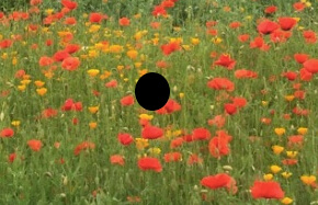
Figure 1
The retinal hole in the center of the macula (arrow) is seen in a color photograph of a patient with a macular hole.
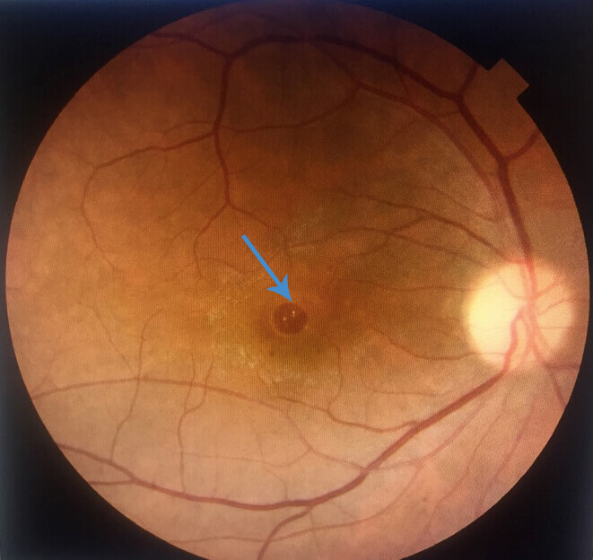
Figure 2
Figure 3:
Huge macular hole (left) closed (right) 2 weeks after surgery.
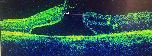
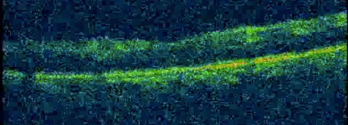
Figure 3
Diabetic retinopathy
Eye involvement is seen among one in three patients with diabetes mellitus. The retina, which is responsible for light perception, is the most commonly affected part of the eye. Retinal involvement is called diabetic retinopathy. Increased blood sugar causes obstruction and increased permeability of the retinal blood vessels. This leads to bleeding and accumulation of fluid in the retina. When the center of the retina, the so-called macula is affected patients complain of blurred and decreased vision. As the disease progresses, new blood vessels appear on the surface of the retina which can easily bleed and cause the retina to detach.
Patients who do not have macular involvement may have no complaints about it critical for them to have an ophthalmic examination at least once a year. Treatment options for diabetic retinopathy are intravitreal injections, laser treatment and a surgical procedure called vitrectomy. Intravitreal injections prevent leakage from the blood vessels and are used to treat for fluid accumulation in the macula. Laser treatment prevents the development of new vessels. Surgical treatment is necessary in patients with bleeding inside the eye and with retinal detachment.
Assoc. Prof. Dr. Sevil Ara Yailali
Figure 1:
Hemorrhage spots and fluid accumulation in the retina are seen in a patient with mild diabetic retinopathy (left). Bleeding in the eye, formation of new blood vessels, and retinal detachment are seen in this image of a patient with severe diabetic retinopathy (middle). The right photo shows a normal retina.
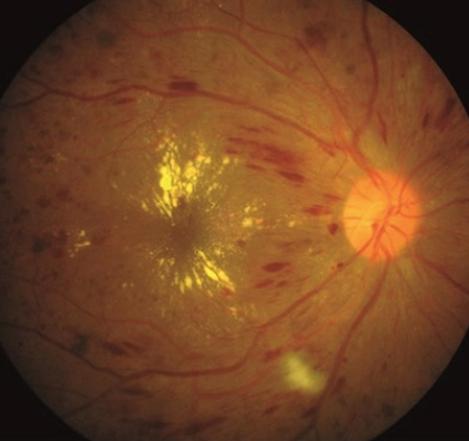
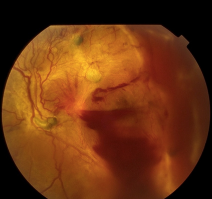
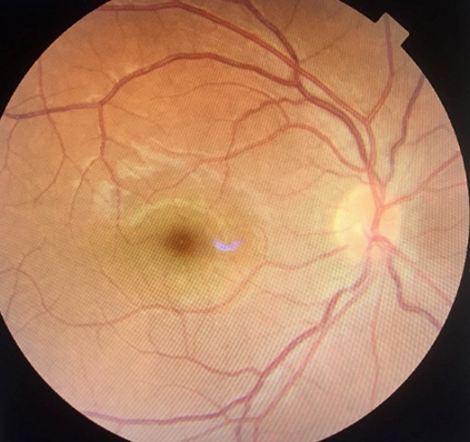
Figure 1
Retinal detachment
Retinal detachment is the separation of the retina from its normal position. The retina is a tissue that is sensitive to light and covers the inner surface at the bottom of the eye. The retina is covered by the vitreous, which fills the cavity of the eyeball and is composed of gel and fluid.
The most common cause of retinal detachment is retinal detachment due to pulling from the vitreous gel. The sudden appearance of clearings and spots in the visual field can be a warning signal of retinal tear.
Entry of the vitreous fluid through the retinal tear site to the space beneath results in its detachment. Signs of retinal detachment may be a decrease in vision or the appearance of a dark spot in the peripheral visual field.
Treatment for retinal tears using a laser procedure can prevent retinal detachment. When retinal detachment occurs, the only treatment is through surgical intervention. Both retinal tear and retinal detachment are emergency conditions, as better visual outcomes are achieved with early treatment.
Assoc. Prof. Dr. Sevil Ara Yailali
Figure 1:
Spots (left) and a dark area (right) on the visual field can be warning signals of a retinal tear or detachment.
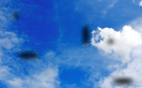

Figure 1
Figure 2:
Pulling on the retina by the vitreous gel (white arrow) causes retinal detachment. The vitreous fluid flowing through the ruptured area (black arrow) to the space under the retina causes the retina to detach.
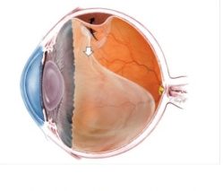
Figure 2
A detached retina (left) is back in place after surgery (right).
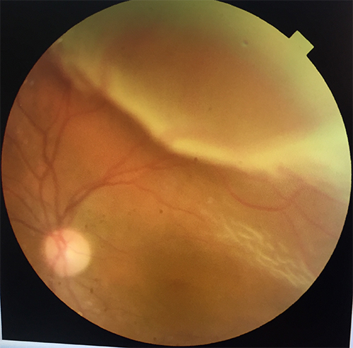
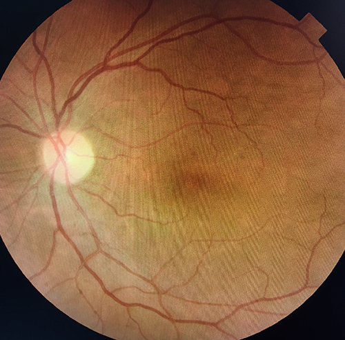
Figure 3