

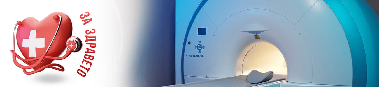
MRI defecography is a specialized type of magnetic resonance imaging that allows specialists to assess how the rectum, anal canal, and pelvic floor function during defecation. The method is completely painless, harmless, and provides extremely accurate information about the anatomy and movements of the organs in the pelvic region.
What is the study?
Unlike standard MRI, which takes static images, dynamic MRI defecography tracks the movement of organs in real time. It helps to identify coordination disorders of muscles and structures involved in defecation.
When is MRI defecography recommended?
The examination may be ordered for patients with the following complaints or suspected diseases:
- Chronic constipation or difficulty with bowel movements
- Feeling of incomplete bowel emptying
- Fecal or gas incontinence (anal incontinence)
Suspicion of:
- Rectal prolapse - when part of the rectum comes out
- Rectocele – bulging of the rectum into the vagina
- Enterocele – herniation of the small intestine into the pelvic cavity
- Pain or pressure in the pelvic area without a clear cause
- Preparation for surgical treatment of the pelvic floor
How does the procedure work?
- Preparation
On the day of the examination, no special diet is required, unless the doctor has recommended otherwise.
A special gel or paste is inserted into the rectum, which mimics the consistency of feces.
The patient changes into hospital attire and is positioned for the examination.
- Positioning
Depending on the equipment, the patient can lie on their back, on their side, or sit up.
Modern systems feature open MRI scanners that provide a more natural position for the examination.
- Filming
Series of images are being made:
- to rest
- When straining
- when attempting to defecate
In some cases, the patient needs to expel the gel introduced during the examination.
- Duration and comfort
- The study usually lasts 20–40 minutes.
- It is not painful, there is no radiation exposure, and it does not require anesthesia.
What does MRI defecography show?
With the help of dynamic imaging, doctors can assess:
- Pelvic floor muscle condition
- Anal sphincter function
- Presence of prolapse, rectocele, enterocele, or other structural changes
- Coordination between the abdominal press and the muscles of the rectal outlet
- Is there a retention of material in the rectum due to a mechanical obstruction or improper muscle contraction?
Advantages of MR defecography
- Painless and non-invasive examination
- No radiation – unlike classic X-ray defecography
- Better visibility of soft tissues – allows simultaneous assessment of the rectum, vagina, bladder, and surrounding muscles
- Dynamic information – shows how organs work in motion, not just their shape
Anorectal manometry defecography is a modern, safe, and highly informative diagnostic method for defecation disorders and pelvic floor dysfunction. It helps doctors identify the exact cause of the problem and choose the most appropriate treatment, whether it's physical therapy, medication, Botox, or surgery.
If you suffer from persistent constipation, a feeling of incomplete bowel movements, or incontinence, consult a gastroenterologist or colorectal surgeon – they can assess whether MRI defecography is appropriate for you.
Medical Karajî Diagnostic and Consultative Center can assist with the organization of MRI defecography by arranging for the examination to be carried out after consultation with the appropriate specialist. This ensures that each patient receives a personalized assessment of their condition and that the examination is tailored to their specific needs and medical history.